


ABSTRACT
Background and Aim: Canine mammary cancer (CMC) is one of the most common malignant neoplasms in female dogs, with high metastatic potential and limited therapeutic options. Natural bioactive compounds derived from medicinal mushrooms have gained increasing attention because of their anticancer properties and low toxicity.
Materials and Methods: The anticancer activity of HE methanolic extract was evaluated using CHMp-13a and CHMp-5b CMC cell lines, while Madin-Darby canine kidney cells were used as normal controls. Cell viability was assessed using the 3-(4,5-dimethyl-2-thiazolyl)-2,5-diphenyl-2H-tetrazolium bromide assay. Cell migration and invasion were evaluated using wound healing and Transwell assays, respectively. Apoptosis was analyzed using Annexin V-fluorescein isothiocyanate/propidium iodide flow cytometry. Relative mRNA and protein expression levels of apoptosis- and EMT-related markers were determined using quantitative real-time polymerase chain reaction and western blotting. The phytochemical profile of the extract was characterized using liquid chromatography quadrupole time-of-flight mass spectrometry.
Results: HE extract significantly inhibited proliferation of both CMC cell lines in a dose- and time-dependent manner, with greater selectivity toward CHMp-13a cells and minimal cytotoxicity in normal cells. Morphological analysis revealed apoptotic features, including cell shrinkage, detachment, and cytoplasmic vacuolization. The extract significantly suppressed migration and invasion capacities of both CMC cell lines. Flow cytometric analysis demonstrated increased apoptotic cell populations following treatment. Molecular analyses showed upregulation of the pro-apoptotic marker BAX and downregulation of the anti-apoptotic marker BCL-2. Furthermore, HE extract suppressed EMT progression by increasing E-cadherin expression while reducing N-cadherin expression. Phytochemical screening identified 17 bioactive compounds, including erinacines, hericenones, hericene derivatives, and phenolic compounds, which may contribute to the observed anticancer activities.
Conclusion: HE extract demonstrated potent
Keywords: apoptosis, canine mammary cancer, epithelial–mesenchymal transition,
INTRODUCTION
The prevalence of age-related diseases in companion animals has increased markedly over the past decade. This trend is largely attributed to improved pet longevity, which consequently increases susceptibility to chronic disorders such as cardiovascular disease, neurodegeneration, and various forms of neoplasia [1, 2]. Among these disorders, neoplastic diseases remain the leading cause of mortality in senior companion animals [3]. In female dogs, mammary gland tumors are the most frequently diagnosed neoplasms worldwide, with approximately 50% of these tumors reported to be malignant [4]. A notably high prevalence of canine mammary tumors has also been documented in Thailand, accounting for 24.5% of all canine biopsy specimens, of which 85.5% were classified as malignant [5]. Canine mammary cancer (CMC) shares several clinical, histopathological, and molecular characteristics with human breast cancer, making it an important comparative model for translational oncology studies [6]. Several factors contribute to the development and progression of CMC, including age, hormonal exposure, genetic predisposition, environmental influences, and nutritional factors [7]. The aggressiveness and clinical outcome of mammary tumors are strongly associated with cellular pleomorphism, mitotic activity, lymphatic invasion, and metastatic dissemination to regional lymph nodes [8].
Cancer progression is characterized by the acquisition of multiple mechanisms that enable tumor cells to survive, proliferate, invade surrounding tissues, and metastasize to distant organs [9]. One of the fundamental hallmarks of cancer involves the evasion of apoptosis, a highly regulated process responsible for maintaining tissue homeostasis through the elimination of damaged or potentially oncogenic cells [10]. Dysregulation of apoptosis-related genes may prevent programmed cell death, thereby facilitating uncontrolled cancer cell proliferation and tumor progression [11]. In particular, decreased expression of the proapoptotic protein BAX together with increased expression of the antiapoptotic protein BCL-2 suppresses apoptotic signaling pathways and promotes cancer cell survival [12]. Another critical mechanism associated with tumor progression is EMT, which enables epithelial cancer cells to acquire migratory and invasive properties [13]. During EMT, epithelial markers such as E-cadherin are downregulated, whereas mesenchymal markers including N-cadherin and SLUG are upregulated, resulting in enhanced motility, invasiveness, and metastatic potential [14, 15]. In CMC, activation of EMT is strongly associated with aggressive tumor behavior and poor prognosis.
Early diagnosis and effective therapeutic intervention are essential for improving survival outcomes and quality of life in dogs affected by mammary cancer. Currently, surgical excision remains the primary treatment strategy for localized mammary tumors, whereas chemotherapeutic agents such as doxorubicin (DOX) and 5-fluorouracil are frequently used in advanced or metastatic cases [16-18]. Nevertheless, conventional chemotherapeutic regimens often exhibit limited efficacy and may induce severe adverse effects, including cardiotoxicity, gastrointestinal disturbances, nephrotoxicity, and myelosuppression in canine patients [19]. Therefore, the development of safer and more effective therapeutic or adjuvant agents capable of suppressing tumor growth and metastasis while minimizing toxicity remains a major challenge in veterinary oncology.
Natural products derived from medicinal mushrooms have attracted considerable scientific attention because of their diverse pharmacological properties, including antioxidant, anti-inflammatory, immunomo-dulatory, antimicrobial, and anticancer activities [20, 21]. Several studies have demonstrated that mushroom-derived bioactive compounds exert antitumor effects through both direct cytotoxic mechanisms and modulation of immune responses [22]. In Asian countries such as Japan and China, medicinal mushrooms are approved and widely used as adjunctive therapies in cancer management, either in combination with chemotherapy and radiotherapy or as supportive single-agent treatments in humans [23]. Consequently, medicinal mushrooms have emerged as promising sources of novel bioactive compounds for pharmaceutical and anticancer drug development [24, 25].
Despite increasing evidence regarding the anticancer properties of HE extract in human cancer models, limited information is available concerning its therapeutic potential in canine cancers, particularly CMC. Most previous studies have focused predominantly on human-derived cancer cell lines, while the biological responses of CMC cells to HE extract remain poorly characterized. In particular, there is insufficient information regarding the effects of HE extract on distinct CMC phenotypes with varying invasive characteristics. Moreover, the molecular mechanisms underlying the anticancer activity of HE extract in CMC have not been comprehensively elucidated. Specifically, there is a lack of evidence regarding its role in regulating apoptosis-associated proteins such as BAX and BCL-2 and EMT-associated markers including E-cadherin, N-cadherin, and SLUG in CMC cells. Furthermore, the phytochemical composition potentially responsible for these biological activities in CMC models remains inadequately characterized. Although several bioactive compounds have previously been identified in HE, their contribution to apoptosis induction, EMT suppression, and antimetastatic activity in CMC cells has not yet been fully investigated. Therefore, additional studies are required to clarify the molecular pathways and phytochemical constituents associated with the anticancer effects of HE extract in CMC models.
Therefore, this study aimed to investigate the
MATERIALS AND METHODS
Ethical approval
This study was conducted entirely under
Study period and location
This study was conducted from December 2024 to December 2025 at the Kasetsart University Animal Cell Bank, Faculty of Veterinary Medicine, Kasetsart University, Bangkok, Thailand. Additional phytochemical analyses were performed in collaboration with specialized analytical laboratories equipped for LC/Q-TOF/MS-based compound characterization.
Study design
This study was designed as an
Mushroom material and extraction
The fruiting bodies of HE were obtained from Organic Sentang Hed Farm Limited Company, Phitsanulok, Thailand. A voucher specimen (BBH No. 50496) was deposited at the Fungarium of BIOTEC Bangkok Herbarium, National Biobank of Thailand, Thailand. The HE fruiting bodies were dried at 60°C and ground into a fine powder. Briefly, 200 g of HE powder was extracted with 95% methanol (MeOH) at a ratio of 10 g/100 mL and stored at 4°C for 24 h. The mixture was subsequently sonicated for 30 min and centrifuged at 10,000 ×
Cell lines and cell culture
The CHMp-13a (low-grade invasive canine mammary adenocarcinoma) and CHMp-5b (high-grade invasive canine mammary adenocarcinoma) cell lines [31] were kindly provided by Professor Dr. Takayuki Nakagawa, Laboratory of Veterinary Surgery, Graduate School of Agricultural and Life Sciences, The University of Tokyo, Japan. The MDCK cell line was obtained from the American Type Culture Collection (ATCC), Manassas, VA, USA.
CMC cells were cultured in RPMI-1640 medium (Corning, New York, NY, USA) supplemented with L-glutamine, 10% fetal bovine serum (FBS; Invitrogen, Carlsbad, CA, USA), and 1% antibiotic-antimycotic solution (Gibco, New York, NY, USA). MDCK cells were maintained in Dulbecco’s Modified Eagle Medium (DMEM; Corning, New York, NY, USA) containing 4.5 g/L glucose and L-glutamine supplemented with 10% FBS and 1% antibiotic-antimycotic solution. All cultures were maintained in a humidified incubator containing 5% CO2 at 37°C. Upon reaching approximately 80% confluence, cells were harvested using 0.25% TrypLE (Gibco) for subculturing.
In this study, CHMp-13a and CHMp-5b cells were used between passages 28 and 41, whereas MDCK cells were used between passages 103 and 107. To ensure experimental integrity and exclude contamination-associated bias, all cell lines were screened for mycoplasma contamination using polymerase chain reaction (PCR)-based detection assays. The results confirmed that all cell lines used in this study were free from mycoplasma contamination (Supplementary Figure S1).
Cell proliferation assay
Cell proliferation was evaluated using the 3-(4,5-dimethyl-2-thiazolyl)-2,5-diphenyl-2H-tetrazolium bromide (MTT) assay according to previously described protocols [32, 33]. Cells were seeded into 96-well plates at a density of 7 × 10³ cells/well and allowed to adhere for 24 h. Subsequently, cells were treated for 48 h with various concentrations of HE extract (0-1000 µg/mL), 0.1% DMSO as the negative control, or DOX at 2 µg/mL as the positive control.
Following treatment, 10 µL of MTT solution was added to each well, and the plates were incubated for an additional 4 h. Thereafter, 100 µL of solubilization buffer was added to each well, followed by overnight incubation at 37°C. The absorbance of the solubilized formazan product was measured at 570 nm using a microplate reader (BioTek, Winooski, VT, USA). All experiments were conducted in quadruplicate across three independent experimental sessions. Blank well absorbance values were subtracted from all readings to eliminate background interference.
Cell viability was expressed as a percentage relative to the negative control group, which was defined as 100% viability, according to the following formula:
Cell viability (%) = [(At − Ab)/(Ac − Ab)] × 100
Where, At = absorbance of treated cells; Ab = absorbance of blank wells; and Ac = absorbance of control cells.
The half-maximal inhibitory concentration (IC50) values were calculated from dose-response curves. In addition, the selectivity index (SI) was used to determine the selective cytotoxicity of HE extract toward cancer cells relative to normal cells using the following equation:
SI = IC50 value (normal cells)/IC50 value (cancer cells)
Cell morphology analysis by phase-contrast microscopy
Based on preliminary proliferation assays and IC50 values, concentrations ranging from 0 to 600 µg/mL were selected for further morphological evaluation of CHMp-13a and CHMp-5b cells. Cells were seeded into 6-well plates at a density of 4 × 10⁵ cells/well and treated with HE extract (0-600 µg/mL). After 48 h of incubation, cellular morphological alterations including changes in cell shape, adherence, shrinkage, and apoptotic features were examined and documented using a phase-contrast microscope (Nikon, Tokyo, Japan) at 10× magnification.
Cell migration assays
Cell migration was initially evaluated using a scratch-wound assay according to previously described methods [32, 33]. Briefly, CMC cells were seeded into 6-well plates at a density of 4 × 10⁵ cells/well and cultured until complete confluence was achieved. A linear wound was generated using a sterile 100 µL pipette tip, after which cells were treated with various concentrations of HE extract (0-600 µg/mL). Wound healing was monitored at 0, 12, and 24 h after scratching. Images were captured using a microscope equipped with NIS-Elements D imaging software version 5.2 (Nikon, Tokyo, Japan). The average wound area was determined using linear measurements obtained from three equidistant positions across each wound. Cell migration was quantified by comparing wound areas at 12 and 24 h relative to the initial wound area at 0 h. Experiments were repeated four times.
The percentage of wound area was calculated as follows:
% wound area = (Wt/W0) × 100
Where, Wt = wound area at time t and W0 = wound area at 0 h.
The migratory capacities of CMC cells were further evaluated using a 24-well Transwell insert system with an 8 µm pore membrane (Corning). CHMp-13a and CHMp-5b cells were suspended in serum-free RPMI-1640 medium and seeded into the upper chamber at a density of 5 × 10³ cells/well. HE extract (0-600 µg/mL) was added to the upper chamber, whereas 0.1% DMSO served as the negative control. The lower chamber contained RPMI-1640 medium supplemented with 10% FBS as a chemoattractant. After 24 h of incubation, nonmigrating cells were removed from the upper chamber, whereas migrated cells attached to the lower membrane surface were fixed with 100% MeOH and stained with Wright-Giemsa stain. Migrated cells were quantified by counting five random microscopic fields per insert, and the average values from three independent experiments were calculated.
Cell apoptosis assay
Apoptosis induction by HE extract was quantified using the Annexin V-FITC apoptosis detection assay (BD Biosciences, San Jose, CA, USA) according to the manufacturer’s instructions. Briefly, 2 × 10⁴ CMC cells/well were seeded into 12-well plates and treated with HE extract at concentrations ranging from 0 to 600 µg/mL for 48 h. Cells treated with 0.1% DMSO served as the negative control, whereas cells treated with DOX (2 µg/mL) served as the positive control.
Following treatment, cells were detached and centrifuged at 400 ×
RNA isolation, cdna synthesis, and qrt-PCR analysis
Total RNA was isolated from CMC cells treated with HE extract (0-600 µg/mL) using the GeneJET RNA Purification Kit (Thermo Fisher Scientific, Waltham, MA, USA) according to the manufacturer’s instructions. Complementary DNA (cDNA) synthesis was subsequently performed using the SuperScript III First-Strand Synthesis System (Invitrogen, Carlsbad, CA, USA) following previously described procedures [32, 33]. Thermal cycling was carried out using a G-Storm GS482 thermal cycler (Gene Technologies, Somerset, UK) under the following conditions: 65°C for 5 min, 50°C for 50 min, and 85°C for 5 min.
Quantitative real-time polymerase chain reaction (qRT-PCR) analysis was performed using a CFX96 Touch Real-Time PCR Detection System (Bio-Rad, Hercules, CA, USA) together with iTaq Universal SYBR Green Supermix (Bio-Rad) according to previously established protocols [33]. The relative mRNA expression levels of
Table 1. Sequences of primers used for quantitative real-time polymerase chain reaction (qRT-PCR).
| Gene | Primer sequence (5′-3′) | Amplicon size (bp) | Accession number | Reference |
|---|---|---|---|---|
|
| F: GGTTGTTGCCCTCCTCTACT R: GTAAGCACTCCAGCCACAAA | 219 | AB080230 | [34] |
|
| F: TGGATGACTGAGTACCTGAA R: GGCCTACTGACTTCACTTAT | 206 | AB116145 | [34] |
|
| F: GGCAAGGCGTTTTCCAGACCCT R: GGGCAAGAAAAAGGCTTCTCCCCAG | 77 | NM_001097981.1 | [35] |
|
| F: TCCTGGGCAGGGTGAGTT R: GAGGCCGCTTGACTGTAATC | 114 | NM_001287125.2 | [36] |
|
| F: AGCACCCTCCTCAGTCAACG R: TGTCAACATGGTCCCAGCA | 128 | NM_001287156.2 | [36] |
|
| F: CCCACTCTTCCACCTTCGAC R: AGCCAAATTCATTGTCATACCAGG | 90 | NM_001003142.2 | [35] |
Western blot analysis
To evaluate the effects of HE extract on protein expression, CMC cells were treated with HE extract at concentrations ranging from 0 to 600 µg/mL for 48 h. Total protein was isolated using ice-cold radioimmunoprecipitation assay (RIPA) buffer (Cell Signaling Technology, Danvers, MA, USA). Cell lysates were sonicated and centrifuged at 14,000 ×
Equal amounts of protein (20 µg) were separated by sodium dodecyl sulfate-polyacrylamide gel electrophoresis (SDS-PAGE) and subsequently transferred onto polyvinylidene difluoride (PVDF) membranes. Membranes were blocked and incubated overnight at 4°C with the following primary antibodies: anti-BAX (#AF0120; 1:2000), anti-BCL-2 (#AF6139; 1:5000), and anti-GAPDH (#AF7021; 1:5000) (Affinity Biosciences, Cincinnati, OH, USA), together with anti-E-cadherin (#14472; 1:2000), anti-N-cadherin (#13116; 1:2000), and anti-SLUG (#95858; 1:2000) (Cell Signaling Technology, Danvers, MA, USA).
After washing with tris-buffered saline containing 0.1% Tween-20, membranes were incubated with horseradish peroxidase (HRP)-conjugated anti-rabbit secondary antibody (#S0001; 1:5000) for 1 h at room temperatura (25°C). Protein bands were visualized using SuperSignal West Pico PLUS Chemiluminescent Substrate (Thermo Fisher Scientific) and detected using a ChemiDoc MP Imaging System (Bio-Rad) with a fixed exposure time of 1 min. Densitometric analysis was performed using Image Lab software (Bio-Rad). Protein expression levels were normalized to GAPDH as the internal loading control and expressed as percentages relative to the untreated control group.
Phytochemical screening by LC/Q-TOF/MS
Phytochemical profiling of HE extract was performed using an LC/Q-TOF/MS system (6546 LC/Q-TOF; Agilent Technologies, Santa Clara, CA, USA) equipped with a Poroshell 120 EC-C18 column (2.7 µm, 3.0 × 150 mm). The analysis was conducted to identify potential bioactive compounds based on molecular mass and chromatographic retention characteristics.
The injection volume was set at 10 µL, with a flow rate of 0.7 mL/min. The column temperature was maintained at 40°C, whereas the autosampler tray temperature was maintained at 10°C throughout the analysis. Gradient elution was performed using a mobile phase consisting of 2 mM ammonium acetate in deuterium-depleted water (mobile phase A) and 100% MeOH (mobile phase B). The gradient program began with 2% mobile phase B for 1 min, gradually increased to 100% within 30 min, and was maintained for 7 min. The system was then returned to initial conditions for 1 min followed by a 3-min equilibration period, resulting in a total run time of 42 min.
Mass spectrometric analysis was conducted in both positive and negative ionization modes. Full-scan mass spectra were collected over an m/z range of 80-1000 at a scan rate of one scan/s.
Statistical analysis
All experimental data are presented as mean ± standard deviation (SD). Statistical analyses for MTT assays were based on three independent experiments, with each treatment performed in quadruplicate. Wound healing assay data were obtained from four independent replicates. Quantitative data generated from Transwell migration assays, apoptosis assays, qRT-PCR analysis, and western blot analysis were derived from three independent experiments.
Data normality was assessed using the Shapiro-Wilk test. Comparisons between HE-treated groups and the control group (0.1% DMSO) were performed using one-way analysis of variance (ANOVA) followed by Dunnett’s multiple comparison test. Differences in IC50 values among cell lines and treatment durations were analyzed using the Extra sum-of-squares F-test. All statistical analyses were performed using GraphPad Prism version 8.0 (GraphPad Software Inc., La Jolla, CA, USA). Statistical significance was considered at p < 0.05.
RESULTS
Cytotoxic effects of HE extract
The cytotoxic effects of HE extract against CMC cell lines and normal MDCK cells were evaluated using the MTT assay. Cells were treated with eight concentrations of HE extract (0-1000 µg/mL) for 24 and 48 h. The dose-response curves of CHMp-13a, CHMp-5b, and MDCK cells following treatment with HE extract are presented as percentages of cell viability in Figure 1. Detailed cell viability data and corresponding statistical analyses for all concentrations and time points are provided in Supplementary Tables S1.
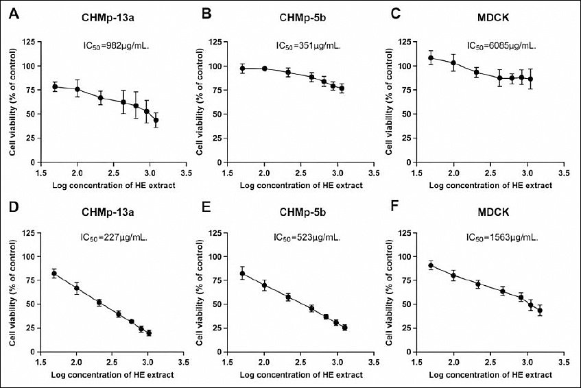
Figure 1. Dose-response curves of CHMp-13a, CHMp-5b, and MDCK cell proliferation following HE extract treatment. Cells were treated with varying concentrations (0-1000 µg/mL) of HE extract for 24 h (A-C) and 48 h (D-F). Data are presented as mean ± SD of three independent experiments.
Following both 24 and 48 h of treatment, HE extract demonstrated significant differences in IC50 values among the tested cell lines (p < 0.0001, Extra sum-of-squares F-test). At both time points, HE extract exhibited greater cytotoxic potency against CHMp-13a cells, with IC50 values of 982 and 227 µg/mL at 24 and 48 h, respectively, compared with CHMp-5b cells, which showed IC50 values of 3510 and 523 µg/mL, respectively. In contrast, minimal cytotoxicity was observed in normal MDCK cells, with IC50 values of 6085 and 1563 µg/mL at 24 and 48 h, respectively (Table 2).
Table 2. IC50 and SI values of HE extract against CHMp-13a, CHMp-5b, and MDCK cells following 24 and 48 h of treatment. Statistical significance was determined using the Extra sum-of-squares F-test.
| Parameter | CHMp-13a | CHMp-5b | MDCK | |||
|---|---|---|---|---|---|---|
|
|
|
| ||||
| 24 h | 48 h | 24 h | 48 h | 24 h | 48 h | |
| IC50 values (µg/mL) | 982 | 227 | 3510 | 523 | 6085 | 1563 |
| 95% confidence interval (95% CI) | 774.9-1356 | 214.4-239.6 | 2750-4770 | 488.9-561 | 3314-16512 | 1329-1910 |
| Selectivity index | 6.19 | 6.92 | 1.73 | 2.98 | - | - |
| p-value | < 0.0001 | < 0.0001 | < 0.0001 | < 0.0001 | - | - |
The SI analysis indicated that an SI value >2 represents selective cytotoxicity toward cancer cells, whereas an SI value <1 indicates greater toxicity toward normal cells. Based on the calculated SI values, HE extract demonstrated selective inhibitory activity against both CMC cell lines. CHMp-13a cells exhibited high selectivity, with SI values of 6.19 and 6.92 at 24 and 48 h, respectively. In comparison, CHMp-5b cells exhibited lower selectivity, with SI values of 1.73 at 24 h and 2.98 at 48 h. These findings indicate that HE extract possesses stronger and more selective antiproliferative activity against CHMp-13a cells than against CHMp-5b cells.
Effect of HE extract on the morphology of CMC cell lines
Morphological alterations in CMC cells following treatment with HE extract were evaluated using phase-contrast microscopy after 48 h of incubation. As shown in Figure 2, HE extract induced concentration-dependent morphological changes in both CHMp-13a and CHMp-5b cells compared with untreated control cells. Microscopic examination demonstrated progressive reductions in cell density together with marked contraction of cell volume as the concentration of HE extract increased.
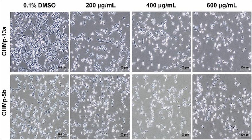
Figure 2. Morphological alterations in CHMp-13a and CHMp-5b cells following treatment with HE extract for 48 h. Concentration-dependent morphological changes were observed following HE extract treatment, including reduced cell population, cytoplasmic vacuolization, cell shrinkage, rounding, and detachment. Images were obtained using phase-contrast inverted microscopy at 10× magnification (scale bar = 100 µm).
At the highest tested concentration (600 µg/mL), both CMC cell lines exhibited prominent cytoplasmic vacuolization, cell shrinkage, cellular rounding, detachment from the culture surface, and floating cells within the culture medium, all of which are characteristic morphological features associated with apoptosis. In contrast, untreated control cells maintained normal cellular morphology and adherence. These observations suggest that HE extract induces marked cytotoxic and apoptotic morphological alterations in CMC cells in a concentration-dependent manner.
HE extract inhibits CMC cell migration
Cancer metastasis is a multistep process involving migration and invasion of tumor cells into surrounding tissues and distant organs. To investigate the antimigratory effects of HE extract, both scratch-wound healing and Transwell migration assays were performed.
The scratch-wound healing assay demonstrated that HE extract significantly inhibited wound closure in both CHMp-13a and CHMp-5b cells following 24 h of treatment (Figure 3). Specifically, treatment with 600 µg/mL HE extract significantly delayed scratch healing in CHMp-13a cells (p = 0.0354) and CHMp-5b cells (p = 0.0347) compared with the untreated control group. Detailed wound area measurements and statistical analyses are provided in Supplementary Tables S2.

Figure 3. Evaluation of CHMp-13a and CHMp-5b cell migration using the wound healing assay following HE extract treatment. (A) Quantitative analysis of wound area in CHMp-13a cells and (C) CHMp-5b cells at 0, 12, and 24 h following treatment with 0-600 µg/mL HE extract. Data are expressed as mean percentage wound area ± SD of four independent replicates. Statistical analysis was performed using one-way ANOVA followed by Dunnett’s multiple comparison test (*p < 0.05 vs. control). (B) Representative wound healing images of CHMp-13a cells and (D) CHMp-5b cells captured at 4× magnification (scale bar = 100 µm).
Similarly, the Transwell migration assay demonstrated a significant concentration-dependent reduction in the migratory capacity of both CMC cell lines following HE extract treatment (Figure 4). In CHMp-13a cells, treatment with 400 and 600 µg/mL HE extract significantly reduced the percentage of migrated cells to 50.65% (p = 0.0002) and 48.46% (p = 0.0001), respectively, relative to the untreated control group (100%). Likewise, in CHMp-5b cells, treatment with 400 and 600 µg/mL HE extract significantly reduced cell migration to 53.45% (p = 0.0003) and 41.42% (p < 0.0001), respectively. Detailed migration cell counts and statistical analyses are provided in Supplementary Tables S3. These findings indicate that HE extract effectively suppresses the migratory behavior of CMC cells in a concentration-dependent manner.

Figure 4. Evaluation of CHMp-13a and CHMp-5b cell migration using the Transwell assay following HE extract treatment. Cells were treated with HE extract (0, 200, 400, and 600 µg/mL) for 24 h. (A, B) Percentages of migrated cells relative to the untreated control are presented as mean ± SD of three independent replicates. Statistical analysis was performed using one-way ANOVA followed by Dunnett’s multiple comparison test (***p < 0.001 and ****p < 0.0001 vs. control). (C) Representative images of migrated cells captured at 4× magnification (scale bar = 100 µm).
HE extract induces apoptosis in CMC cells
To determine whether HE extract induces apoptosis in CMC cells, Annexin V-FITC/PI double staining followed by flow cytometric analysis was performed. Cell populations were classified according to staining characteristics as viable cells, necrotic cells, early apoptotic cells, and late apoptotic cells.
Treatment with HE extract (0-600 µg/mL) for 48 h induced a significant concentration-dependent increase in apoptotic cell populations in both CHMp-13a and CHMp-5b cells (Figure 5A). In CHMp-13a cells, treatment with 600 µg/mL HE extract significantly increased the percentages of early apoptotic and late apoptotic cells from 1.49% and 1.72% in untreated controls to 3.65% (p = 0.0011) and 17.97% (p = 0.0002), respectively (Figure 5B and 5C). Similarly, treatment of CHMp-5b cells with 600 µg/mL HE extract significantly increased late apoptotic cells from 2.00% in the untreated control group to 7.94% (p < 0.0001) (Figure 5C). Detailed apoptotic cell data and corresponding statistical analyses are provided in Supplementary Tables S4.
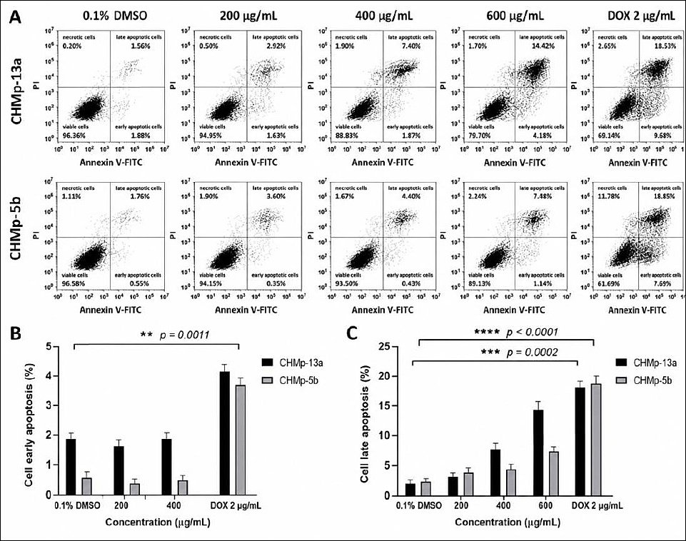
Figure 5. Flow cytometric quantification of apoptotic CHMp-13a and CHMp-5b cells following HE extract treatment. (A) Representative flow cytometry scatter plots showing Annexin V-FITC and PI staining after 48 h of treatment. (B) Percentages of early apoptotic cells (Annexin V+/PI−) and (C) late apoptotic cells (Annexin V+/PI+) relative to the total cell population. Data are presented as mean ± SD of three independent replicates. Statistical significance was determined relative to the untreated control group (**p < 0.01, ***p < 0.001, and ****p < 0.0001). DOX (2 µg/mL) and 0.1% DMSO served as positive and negative controls, respectively.
These findings demonstrate that HE extract effectively induces apoptosis in both CMC cell lines in a concentration-dependent manner.
Relative mrna expression levels in CMC cells
To elucidate the molecular mechanisms underlying HE extract-induced apoptosis and EMT modulation, the relative mRNA expression levels of apoptosis- and EMT-associated markers were quantified in both CMC cell lines using qRT-PCR analysis (Figures 6A and 6B).
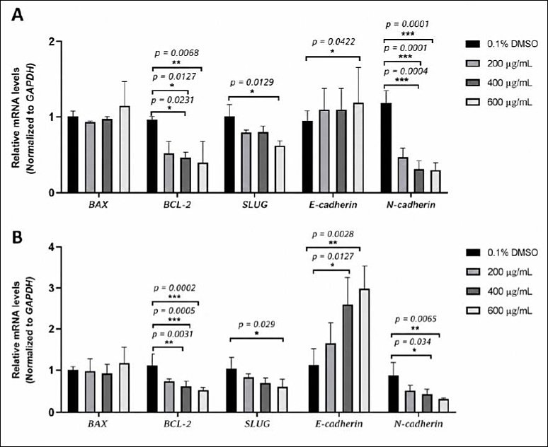
Figure 6. Relative mRNA expression levels in HE-treated CHMp-13a and CHMp-5b cells. Effects of HE extract on the expression of
In CHMp-13a cells, HE extract significantly reduced
EMT is widely recognized as a critical biological process associated with enhanced invasiveness and metastatic dissemination of cancer cells [37]. A hallmark feature of EMT is the “cadherin switch,” characterized by reduced E-cadherin expression and increased N-cadherin expression, resulting in loss of intercellular adhesion and enhanced cellular motility [38]. To further investigate the inhibitory effects of HE extract on EMT, the expression levels of
Following treatment with HE extract, a marked shift in EMT-related gene expression profiles was observed. HE extract significantly increased
Conversely, expression of the mesenchymal markers
Collectively, these findings suggest that HE extract may interfere with EMT progression in CMC cells by enhancing epithelial marker expression while suppressing mesenchymal-associated markers. These molecular alterations indicate the potential antimetastatic activity of HE extract in CMC cells. Further studies are warranted to elucidate the precise signaling pathways underlying the apoptosis-inducing and EMT-suppressive effects of HE extract.
Protein expression by western blot analysis
To further evaluate the molecular effects of HE extract on apoptosis- and EMT-related pathways, western blot analysis was performed to determine the protein expression levels of BAX, BCL-2, E-cadherin, N-cadherin, and SLUG in both CMC cell lines following 48 h of treatment with HE extract.
Alterations in BCL-2 family proteins play essential roles in regulating apoptosis [39]. In CHMp-13a cells (Figures 7A and 7B), HE extract significantly increased BAX protein expression at 400 µg/mL (p = 0.0016) and 600 µg/mL (p < 0.0001), whereas BCL-2 protein expression was significantly decreased at the same concentrations (p = 0.0385 and p = 0.0035, respectively). Similarly, in CHMp-5b cells (Figures 8A and 8B), HE extract significantly elevated BAX protein expression at 400 µg/mL (p = 0.0005) and 600 µg/mL (p < 0.0001), accompanied by significant reductions in BCL-2 expression at 400 µg/mL (p = 0.0306) and 600 µg/mL (p = 0.0003). These findings support the involvement of HE extract in promoting apoptosis through modulation of intrinsic apoptotic pathways.
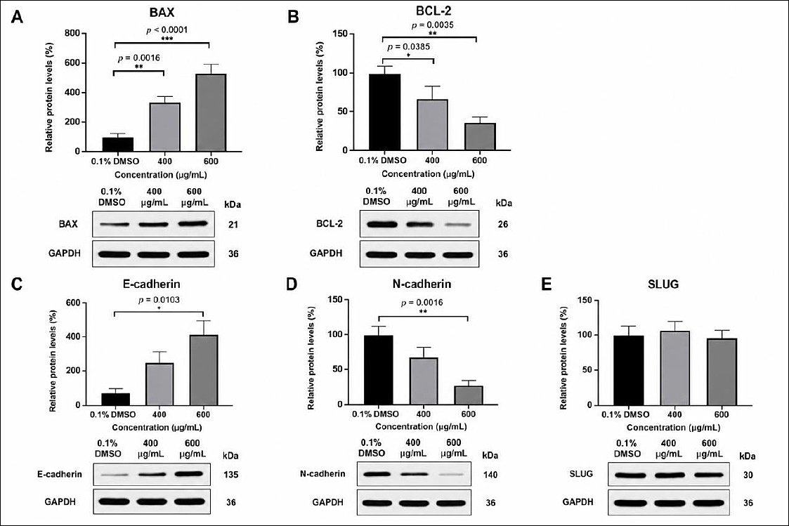
Figure 7. Western blot analysis of BAX, BCL-2, E-cadherin, N-cadherin, and SLUG proteins in CHMp-13a cells following HE extract treatment. (A-E) Cells were treated with 0, 400, and 600 µg/mL HE extract for 48 h. Protein expression levels were normalized to GAPDH expression. Data are presented as mean ± SD of three independent replicates. Statistical analyses were performed using one-way ANOVA followed by Dunnett’s multiple comparison test against the control group (0.1% DMSO). Statistical significance was considered at *p <0.05, **p < 0.01, ***p < 0.001, and ****p<0.0001.
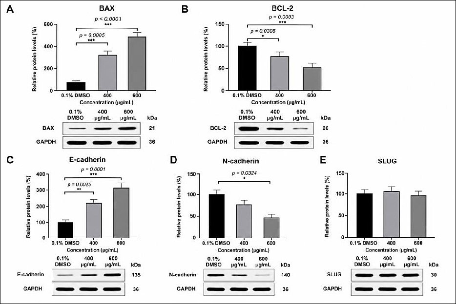
Figure 8. Western blot analysis of BAX, BCL-2, E-cadherin, N-cadherin, and SLUG proteins in CHMp-5b cells following HE extract treatment. (A-E) Cells were treated with 0, 400, and 600 µg/mL HE extract for 48 h. Protein expression levels were normalized to GAPDH expression. Data are presented as mean ± SD of three independent replicates. Statistical analyses were performed using one-way ANOVA followed by Dunnett’s multiple comparison test against the control group (0.1% DMSO). Statistical significance was considered at *p <0.05, **p < 0.01, ***p < 0.001, and ****p<0.0001.
To further investigate the role of HE extract in suppressing metastatic potential, the expression of EMT-associated proteins was also examined. In CHMp-13a cells, E-cadherin expression was significantly increased at 600 µg/mL (p = 0.0103), whereas N-cadherin expression was significantly reduced at the same concentration (p = 0.0016) (Figures 7C and 7D). Similarly, in CHMp-5b cells, E-cadherin expression significantly increased at 400 µg/mL (p = 0.0025) and 600 µg/mL (p = 0.0001), whereas N-cadherin expression was significantly decreased at 600 µg/mL (p = 0.0324) (Figures 8C and 8D).
In contrast, no significant alterations in SLUG protein expression were detected in either cell line at any tested concentration (Figures 7E and 8E). Detailed protein expression data, statistical analyses, and uncropped western blot images are presented in Supplementary Tables S6 and Supplementary Figure S3. Overall, these findings suggest that HE extract modulates the metastatic phenotype of CMC cells through suppression of EMT-associated molecular alterations together with regulation of intrinsic apoptotic signaling pathways mediated by BCL-2 family proteins.
Phytochemical screening analysis
Phytochemical characterization of the methanolic HE extract using LC/Q-TOF/MS in both positive and negative ionization modes revealed the presence of multiple bioactive compounds. A total of 17 phytochemical compounds with potential anticancer activity were identified based on retention time (Rt), peak intensity, and molecular mass analysis (Table 3 and Supplementary Figure S4).
Table 3. Identified phytochemical compounds in HE extract determined by liquid chromatography quadrupole time-of-flight mass spectrometry (LC/Q-TOF/MS).
| No. | Identified compounds | Rt (min) | Formula | [M+H]+ (m/z) | [M+H]− (m/z) | MS/MS fragment ion (m/z) |
|---|---|---|---|---|---|---|
| 1 | Herierin IV | 5.722 | C8H10O4 | 170.0584 | - | 171.0657, 172.0688, 193.0475 |
| 2 | Herniarin | 12.950 | C10H8O3 | 177.0546 | - | 177.0554, 194.0821, 199.0366 |
| 3 | Gingerenone B | 13.008 | C22H26O6 | 386.1739 | - | 387.1802, 404.2068, 409.1622 |
| 4 | Erinacerin G | 16.542 | C16H19NO5 | 305.1292 | - | 306.1336, 323.1601, 328.1155 |
| 5 | Coriandrone E | 18.515 | C13H12O5 | 248.0689 | - | 249.0757, 266.1023, 271.0577 |
| 6 | Calomelanol J | 20.253 | C24H18O5 | 386.1139 | - | 387.1227, 404.1492, 409.1046 |
| 7 | 4-Gingerol | 20.600 | C15H22O4 | 266.1523 | - | 267.1591, 284.1856, 289.1410 |
| 8 | Uncarine F | 21.328 | C21H24N2O4 | 368.1741 | - | 369.1809, 386.2074, 391.1628 |
| 9 | Garcinone E | 22.920 | C28H32O6 | 464.2217 | - | 465.2272, 466.2306, 482.2537, 483.2570 |
| 10 | Erinacine A | 25.888 | C25H36O6 | 432.2532 | - | 433.2585, 434.2619, 450.2850, 451.2883 |
| 11 | Hericenone B | 27.661 | C27H31NO4 | 433.2267 | - | 434.2326, 435.2359, 451.2591, 452.2624 |
| 12 | 8-Gingerol | 28.033 | C19H30O4 | 322.2146 | - | 323.2217, 340.2482, 345.2036 |
| 13 | Hericerin | 30.593 | C27H33NO3 | 419.2468 | - | 420.2533, 421.2566, 438.2831 |
| 14 | Hericenone F | 31.087 | C35H54O6 | 570.3954 | - | 571.3993, 572.4027, 588.4259, 589.4292 |
| 15 | Sesaminol 2-O-triglucoside | 31.989 | C36H46O22 | 830.2511 | - | 831.2588, 848.2819, 849.2853 |
| 16 | Hericene A | 33.319 | C35H56O5 | - | 556.4106 | 557.4201, 558.4235, 574.4466, 575.4500 |
| 17 | Hericene B | 34.587 | C37H58O5 | - | 582.4294 | 583.4357, 584.4391, 600.4623, 601.4656 |
Among the identified compounds, hericenones and erinacines represented the major classes of monoterpenoid compounds. These compounds have previously been associated with diverse biological activities, including antioxidant, anticancer, neuroprotective, immunoregulatory, antibacterial, and hypoglycemic effects [40]. In addition, hericene and hericerin, which are classified as geranyl resorcinol meroterpenoids predominantly identified in
Several additional bioactive constituents, including phenolic compounds, polyphenols, pyranones, terpenoid alkaloids, xanthones, benzaldehydes, and oligosaccharides, were also identified in HE extract. The presence of these diverse phytochemicals suggests that HE extract possesses a complex phytochemical composition that may contribute synergistically to its observed anticancer activities. These findings further support the potential therapeutic application of HE extract as a source of bioactive compounds for future functional and mechanistic anticancer studies.
DISCUSSION
Therapeutic challenges in CMC
Mammary cancer is one of the most frequently diagnosed neoplastic diseases in women and female dogs and remains a major public health and veterinary concern worldwide. CMC is a multifactorial disease influenced by several genetic, hormonal, environmental, and nutritional factors that contribute to tumor initiation and progression [42]. As one of the most prevalent cancers in dogs, CMC markedly affects survival rate and quality of life, thereby creating substantial therapeutic challenges for veterinary clinicians. Surgical excision remains the principal treatment modality for localized mammary tumors and commonly involves removal of the tumor together with regional lymph node dissection to minimize metastatic spread. In advanced or metastatic cases, chemotherapy is often administered as adjuvant or palliative therapy following surgery to reduce recurrence and improve overall survival [43, 44]. Nevertheless, conventional chemotherapeutic regimens are frequently associated with adverse side effects and variable therapeutic outcomes. Consequently, increasing attention has been directed toward the identification of alternative therapeutic agents or adjuvant strategies capable of enhancing treatment efficacy while minimizing toxicity and improving patient quality of life.
Anticancer potential of medicinal mushrooms and HE extract
Medicinal mushrooms possess diverse biological activities, including antimicrobial, anticancer, immuno-modulatory, antioxidant, anti-inflammatory, antidiabetic, and prebiotic properties [45, 46]. Importantly, these natural products are generally associated with low toxicity and minimal adverse effects, making them attractive candidates for novel therapeutic development [47, 48]. Numerous studies have demonstrated that mushroom-derived compounds such as phenolic acids, polyphenols, flavonoids, polysaccharides, and terpenoids modulate critical molecular pathways involved in cancer initiation, progression, apoptosis, metastasis, and oxidative stress regulation [49, 50]. For example, Li
Selective antiproliferative activity of HE extract
In the present study, HE extract demonstrated potent antiproliferative activity against both CHMp-13a and CHMp-5b CMC cell lines, with greater sensitivity observed in CHMp-13a cells. Following 48 h of treatment, the IC50 values were 227 µg/mL for CHMp-13a cells and 523 µg/mL for CHMp-5b cells. In contrast, normal MDCK cells exhibited substantially higher IC50 values (1563 µg/mL), indicating relatively low cytotoxicity toward noncancerous cells. Moreover, SI values of 6.92 for CHMp-13a cells and 2.98 for CHMp-5b cells further confirmed the selective cytotoxic activity of HE extract toward malignant cells while sparing normal cells at therapeutic concentrations. These findings highlight the potential application of HE extract as a selective therapeutic or adjuvant agent in canine mammary oncology.
The observed IC50 values obtained in this study are consistent with previous findings in human breast cancer models. Atay
Inhibition of CMC cell migration and EMT modulation
Metastasis is a critical step in cancer progression and involves migration and invasion of tumor cells from the primary site to distant organs [62]. Therefore, inhibition of tumor migration represents an important therapeutic target for limiting metastatic dissemination. In the present study, both scratch-wound healing and Transwell migration assays demonstrated that HE extract significantly suppressed migration of CHMp-13a and CHMp-5b cells in a concentration-dependent manner. Treatment with 400 µg/mL HE extract reduced migration by approximately 50%, whereas 600 µg/mL markedly delayed wound closure and significantly inhibited cell migration in both CMC cell lines. These findings indicate that HE extract possesses substantial antimetastatic activity against CMC cells.
EMT is widely recognized as a major biological mechanism contributing to cancer metastasis and tumor aggressiveness [63]. This process is characterized by downregulation of epithelial markers such as E-cadherin and upregulation of mesenchymal markers including N-cadherin and SLUG, leading to loss of cell-cell adhesion and enhanced cellular motility [64]. In this study, HE extract significantly increased E-cadherin expression while simultaneously suppressing N-cadherin expression at both mRNA and protein levels in CHMp-13a and CHMp-5b cells. Furthermore, significant reductions in
Apoptosis induction through BAX/BCL-2 regulation
Induction of apoptosis is one of the principal strategies in cancer therapy for preventing tumor progression, recurrence, and metastasis [67]. Morphological characteristics of apoptosis include cell rounding, shrinkage, detachment, cytoplasmic vacuolization, and loss of intercellular adhesion [68, 69]. In the present study, HE extract induced distinct apoptotic morphological alterations in both CMC cell lines, including cytoplasmic vacuolization, cell shrinkage, rounding, and reduced cell-cell adhesion. These findings were further supported by Annexin V-FITC/PI flow cytometric analysis, which demonstrated significant increases in apoptotic cell populations following treatment with HE extract. Treatment with 600 µg/mL HE extract for 48 h significantly increased late apoptotic cell populations in both CHMp-13a and CHMp-5b cells compared with the 0.1% DMSO control group. These observations indicate that HE extract effectively induces apoptosis in CMC cells.
Mitochondrial-mediated apoptosis represents the predominant form of programmed cell death in mammalian cells and is tightly regulated by members of the BCL-2 family proteins [70]. Proapoptotic proteins include BAX, Bcl-2 homologous antagonist/killer, Bcl-2-related ovarian killer, and Bcl-2-associated agonist of cell death, whereas antiapoptotic proteins include BCL-2, B-cell lymphoma-w, and B-cell lymphoma-extra-large [71]. In the present study, HE extract significantly increased BAX expression while simultaneously decreasing BCL-2 expression at both mRNA and protein levels in both CMC cell lines. These findings suggest that HE extract induces apoptosis primarily through modulation of the mitochondrial apoptotic pathway by shifting the BAX/BCL-2 ratio toward a proapoptotic state. Similar observations have previously been reported in human U937 leukemia cells, where HE extract activated mitochondria-mediated apoptotic signaling and increased the ratio of proapoptotic to antiapoptotic proteins [49]. Nevertheless, additional mechanistic studies involving caspase activation assays and ROS quantification are required to further distinguish the involvement of intrinsic and extrinsic apoptotic pathways in HE-treated CMC cells.
Phytochemical contribution to anticancer activity
The anticancer activity of HE extract observed in this study may be attributed to its diverse phytochemical composition, including terpenoids, phenolic compounds, flavonoids, alkaloids, and polysaccharides, which are known to exert antioxidant, cytotoxic, immunomodulatory, and antimetastatic effects [72]. In particular, erinacines and hericenones are considered the major therapeutically active compounds in HE mushrooms and have previously been shown to regulate mitochondrial apoptosis through modulation of the BAX/BCL-2 ratio in colorectal cancer cells. Erinacine A has been reported to activate both intrinsic and extrinsic apoptotic pathways in DLD-1 and HCT-116 colorectal cancer cells [73]. Furthermore, Ruan
Strengths, limitations, and future perspectives
Previous toxicological investigations further support the safety profile of HE extracts. Studies evaluating aqueous and methanolic HE extracts in isolated mouse hepatocytes reported minimal cytotoxicity [78]. Furthermore, long-term toxicity studies demonstrated that HE powder produced no adverse effects in rats even at doses of 2000 mg/kg administered over 90 days [79]. These findings suggest that HE extract may represent a promising candidate for development as a potential adjuvant therapeutic agent for CMC management.
One of the major strengths of the present study is the comprehensive evaluation of HE extract using multiple biological approaches, including antiproliferative assays, migration assays, apoptosis analysis, EMT-associated molecular evaluation, western blot analysis, and phytochemical profiling. Additionally, the use of two distinct CMC cell lines with different invasive phenotypes provides broader insight into the therapeutic potential of HE extract in canine mammary oncology. The integration of LC/Q-TOF/MS phytochemical characterization further strengthens the translational relevance of the study by identifying multiple bioactive compounds potentially associated with the observed anticancer activities.
However, several limitations should also be acknowledged. First, the present study was conducted entirely under
Therefore, future investigations should focus on standardization of HE extract through identification and quantification of key bioactive compounds, evaluation of ROS generation and caspase activation, assessment of potential synergistic interactions with conventional chemotherapeutic agents such as DOX, and
CONCLUSION
This study demonstrated that HE extract possesses significant
The phytochemical profiling by LC/Q-TOF/MS identified multiple bioactive compounds, including erinacines, hericenones, hericene A, and hericerin, which may collectively contribute to the observed anticancer activities. These findings highlight the therapeutic potential of HE extract as a promising natural source of bioactive compounds for CMC management. A major strength of this study was the integrated evaluation of antiproliferative activity, apoptosis induction, EMT modulation, protein expression, and phytochemical characterization using two CMC cell lines with distinct invasive phenotypes.
Nevertheless, this study also has several limitations. The experiments were conducted entirely under
Future studies should focus on
Collectively, the findings of this study suggest that HE extract may serve as a promising candidate for development as an adjuvant therapeutic agent for canine mammary cancer by promoting apoptosis and suppressing EMT-associated migration in CMC cells.
DATA AVAILABILITY
The supplementary data can be made available from the corresponding author upon request.
AUTHORS’ CONTRIBUTIONS
UJ: Conceptualization, methodology, software, validation, formal analysis, investigation, data curation, writing – original draft preparation, writing – review and editing, and project administration. AS: Methodology, formal analysis, validation, data curation, and writing – original draft preparation. TJ: Supervision, methodology, and writing – original draft preparation. SK: Conceptualization, supervision, and visualization. SS: Methodology and writing – review and editing. WS: Methodology and writing – review and editing. KS: Supervision, visualization, and data curation. AK: Investigation and writing – original draft preparation. YBY: Methodology, formal analysis, investigation, data curation, and writing – original draft preparation. All authors have read and approved the final version of the manuscript.
COMPETING INTERESTS
The authors declare that they have no competing interests.
PUBLISHER’S NOTE
Veterinary World remains neutral with regard to jurisdictional claims in the published institutional affiliations.
ACKNOWLEDGMENTS
This research was funded by the Kasetsart Veterinary Development Funds, Faculty of Veterinary Medicine, Kasetsart University, Bangkok, Thailand, under grant number VET.KU.62_02. The authors sincerely thank Professor Dr. Takayuki Nakagawa, Laboratory of Veterinary Surgery, Graduate School of Agricultural and Life Sciences, The University of Tokyo, Japan, for generously providing the CHMp cell lines used in this study. The authors also extend their appreciation to the Laboratory of Toxicology, Department of Environmental Veterinary Science, Faculty of Veterinary Medicine, Hokkaido University, Japan, for their expert assistance with the phytochemical screening analysis.
REFERENCES
- Sarver AL, Makielski KM, DePauw TA, Schulte AJ, Modiano JF. Increased risk of cancer in dogs and humans: A consequence of recent extension of lifespan beyond evolutionarily-determined limitations?. Aging Cancer 2022;3(1):3-19. [Google Scholar] | [Crossref]
- Tanaka M, Yamaguchi S, Iwasa Y. Enhanced risk of cancer in companion animals as a response to the longevity. Sci Rep 2020;10(1):19508. [Google Scholar] | [Crossref]
- Fonti N, Parisi F, Lachi A, Dhein ES, Guscetti F, Poli A. Age at tumor diagnosis in 14,636 canine cases from the pathology-based UNIPI animal cancer registry, Italy: One size doesn't fit all. Vet Sci 2024;11(10):485. [Google Scholar] | [Crossref]
- Pinho SS, Carvalho S, Cabral J, Reis CA, Gärtner F. Canine tumors: A spontaneous animal model of human carcinogenesis. Transl Res 2012;159(3):165-72. [Google Scholar] | [Crossref]
- Srisawat W, Pringproa K, Prachasilchai W, Thongtharb A, Sthitmatee N. Epidemiology and classification for canine and feline mammary gland tumors: A histopathological survey of 437 mammary gland tumor biopsies performed in a secondary care hospital in Chiang Mai, Thailand from 2012 to 2019. PeerJ 2024;12:e17077. [Google Scholar] | [Crossref]
- Kwon JY, Moskwa N, Kang W, Fan TM, Lee C. Canine as a comparative and translational model for human mammary tumor. J Breast Cancer 2023;26(1):1. [Google Scholar] | [Crossref]
- Vazquez E, Lipovka Y, Cervantes-Arias A, Garibay-Escobar A, Haby MM, Queiroga FL. Canine mammary cancer: State of the art and future perspectives. Animals 2023;13(19). [Google Scholar] | [Crossref]
- Goldschmidt M, Peña L, Rasotto R, Zappulli V. Classification and grading of canine mammary tumors. Vet Pathol 2011;48(1):117-31. [Google Scholar] | [Crossref]
- Liu Z, Chen J, Ren Y, Liu S, Ba Y, Zuo A. Multi-stage mechanisms of tumor metastasis and therapeutic strategies. Signal Transduct Target Ther 2024;9(1):270. [Google Scholar] | [Crossref]
- Morana O, Wood W, Gregory CD. The apoptosis paradox in cancer. Int J Mol Sci 2022;23(3):1328. [Google Scholar] | [Crossref]
- He R, Liu Y, Fu W, He X, Liu S, Xiao D. Mechanisms and cross-talk of regulated cell death and their epigenetic modifications in tumor progression. Mol Cancer 2024;23(1):267. [Google Scholar] | [Crossref]
- Lopez A, Reyna DE, Gitego N, Kopp F, Zhou H, Miranda-Roman MA. Co-targeting of BAX and BCL-XL proteins broadly overcomes resistance to apoptosis in cancer. Nat Commun 2022;13(1):1199. [Google Scholar] | [Crossref]
- Tiwari N, Gheldof A, Tatari M, Christofori G. EMT as the ultimate survival mechanism of cancer cells. Semin Cancer Biol 2012;22(3):194-207. [Google Scholar] | [Crossref]
- Varallo GR, Gelaleti GB, Maschio-Signorini LB, Moschetta MG, Lopes JR, De Nardi AB. Prognostic phenotypic classification for canine mammary tumors. Oncol Lett 2019;18(6):6545-53. [Google Scholar] | [Crossref]
- Loh CY, Chai JY, Tang TF, Wong WF, Sethi G, Shanmugam MK. The E-cadherin and N-cadherin switch in epithelial-to-mesenchymal transition: signaling, therapeutic implications, and challenges. Cells 2019;8(10):1118. [Google Scholar] | [Crossref]
- Kuruoglu FE, Ekici ZM, Nak D, Ozyigit MO, Kupeli ZA, Koca D. Investigation of efficacy of two different chemotherapy protocols used in neoadjuvant chemotherapy in clinical stages II-IV canine malignant mammary tumours. Vet Comp Oncol 2024;22(2):284-94. [Google Scholar] | [Crossref]
- Kim Y, Seo K, Song K. Chemotherapy of mammary comedocarcinoma with doxorubicin in a dog. J Vet Clin 2021;38(6):274-8. [Google Scholar] | [Crossref]
- Karayannopoulou M, Kaldrymidou E, Constantinidis TC, Dessiris A. Adjuvant post-operative chemotherapy in bitches with mammary cancer. J Vet Med A Physiol Pathol Clin Med 2001;48(2):85-96. [Google Scholar] | [Crossref]
- Silva LP, Yamamoto PA, Machado MCA, Neves FMF, Azeredo FJ, dos Santos Silva ACS. A pilot study of chemotherapy combinations in rats: Focus on mammary cancer treatment in female dogs. Res Vet Sci 2023;156:14-21. [Google Scholar] | [Crossref]
- Figueiredo L, Régis WCB. Medicinal mushrooms in adjuvant cancer therapies: An approach to anticancer effects and presumed mechanisms of action. Nutrire 2017;42(1):28. [Google Scholar] | [Crossref]
- Sadowska A, Włosek-Pawełas D, Car H. Medicinal mushrooms and their bioactive compounds: From traditional use to therapeutic potential. Molecules 2026;31(10):1749. [Google Scholar] | [Crossref]
- Pathak MP, Pathak K, Saikia R, Gogoi U, Ahmad MZ, Patowary P. Immunomodulatory effect of mushrooms and their bioactive compounds in cancer: A comprehensive review. Biomed Pharmacother 2022;149:112901. [Google Scholar] | [Crossref]
- Rossi P, Difrancia R, Quagliariello V, Savino E, Tralongo P, Lucia Randazzo C. β-glucans from
Grifola frondosa andGanoderma lucidum in breast cancer: An example of complementary and integrative medicine. Oncotarget 2018;9(37). [Google Scholar] | [Crossref] - Cateni F, Gargano ML, Procida G, Venturella G, Cirlincione F, Ferraro V. Mycochemicals in wild and cultivated mushrooms: Nutrition and health. Phytochem Rev 2022;21(2):339-83. [Google Scholar] | [Crossref]
- Kozarski M, Klaus A, van Griensven L, Jakovljevic D, Todorovic N, Wan WAAQI. Mushroom β-glucan and polyphenol formulations as natural immunity boosters and balancers: Nature of the application. Food Sci Hum Wellness 2023;12(2):378-96. [Google Scholar] | [Crossref]
- Gravina AG, Pellegrino R, Auletta S, Palladino G, Brandimarte G, D'Onofrio R.
Hericium erinaceus , a medicinal fungus with a centuries-old history: Evidence in gastrointestinal diseases. World J Gastroenterol 2023;29(20):3048-65. [Google Scholar] | [Crossref] - Tan YF, Mo JS, Wang YK, Zhang W, Jiang YP, Xu KP. The ethnopharmacology, phytochemistry and pharmacology of the genus
Hericium . J Ethnopharmacol 2024;319:117353. [Google Scholar] | [Crossref] - Atmaca H, Çamli Pulat Ç, Ilhan S, Kalyoncu F.
Hericium erinaceus extract induces apoptosis via PI3K/AKT and RAS/MAPK signaling pathways in prostate cancer cells. Chem Biodivers 2024;21(12):e202400905. [Google Scholar] | [Crossref] - Rüstem DG, Aydin HH, Kalmis E, Kayalar H, Ak H. The effects of
Hericium erinaceus extracts on cell viability and telomerase activity in MCF-7 cells. Turk J Biochem 2023;48(3):298-302. [Google Scholar] | [Crossref] - Atay S, Ak H, Kalmis E, Kayalar H, Aydin H. Transcriptome wide analysis reveals the molecular mechanism of tumoricidal effects of lion's mane medicinal mushroom,
Hericium erinaceus (Agaricomycetes) on MCF-7 breast cancer cells. Int J Med Mushrooms 2020;23. [Google Scholar] | [Crossref] - Murai K, Nakagawa T, Endo Y, Kamida A, Yoshida K, Mochizuki M. Establishment of a pair of novel cloned tumour cell lines with or without metastatic potential from canine mammary adenocarcinoma. Res Vet Sci 2012;93(1):468-72. [Google Scholar] | [Crossref]
- Jianpraphat N, Supsavhad W, Ngernmeesri P, Siripattarapravat K, Soontararak S, Akrimajirachoote N. A new benzo[6,7]oxepino[3,2-b] pyridine derivative induces apoptosis in canine mammary cancer cell lines. Animals 2024;14(3). [Google Scholar] | [Crossref]
- Jermnak U, Supsavhad W, Kunakornsawat S, Jaroensong T, Watcharasit P, Visitnonthachai D. Anti-cancer potentials of
Gynura procumbens leaves extract against two canine mammary cancer cell lines. Vet Med Sci 2022;8(1):69-84. [Google Scholar] | [Crossref] - Panja K, Buranapraditkun S, Roytrakul S, Kovitvadhi A, Lertwatcharasarakul P, Nakagawa T. Scorpion venom peptide effects on inhibiting proliferation and inducing apoptosis in canine mammary gland tumor cell lines. Animals 2021;11(7). [Google Scholar] | [Crossref]
- Elshafae SM, Hassan BB, Supsavhad W, Dirksen WP, Camiener RY, Ding H. Gastrin-releasing peptide receptor (GRPr) promotes EMT, growth, and invasion in canine prostate cancer. Prostate 2016;76(9):796-809. [Google Scholar] | [Crossref]
- Yu C, Zheng H, Liu X, Xie G. The analysis of E-cadherin, N-cadherin, vimentin, HER-2, CEA, CA15-3 and SF expression in the diagnosis of canine mammary tumors. Animals 2022;12(21). [Google Scholar] | [Crossref]
- Sciacovelli M, Frezza C. Metabolic reprogramming and epithelial-to-mesenchymal transition in cancer. FEBS J 2017;284(19):3132-44. [Google Scholar] | [Crossref]
- Gheldof A, Berx G. Cadherins and epithelial-to-mesenchymal transition. Prog Mol Biol Transl Sci 2013;116:317-36. [Google Scholar] | [Crossref]
- Qian S, Wei Z, Yang W, Huang J, Yang Y, Wang J. The role of BCL-2 family proteins in regulating apoptosis and cancer therapy. Front Oncol 2022;12:985363. [Google Scholar] | [Crossref]
- Qiu Y, Lin G, Liu W, Zhang F, Linhardt RJ, Wang X. Bioactive compounds in
Hericium erinaceus and their biological properties: A review. Food Sci Hum Wellness 2024;13(4):1825-44. [Google Scholar] | [Crossref] - Kobayashi S. Total synthesis of geranyl-resorcinols isolated from mushrooms of genus
Hericium . Synthesis 2022;55(03):417-32. [Google Scholar] | [Crossref] - Sorenmo K, Rasotto R, Zappulli V, Goldschmidt M. Development, anatomy, histology, lymphatic drainage, clinical features, and cell differentiation markers of canine mammary gland neoplasms. Vet Pathol 2011;48(1):85-97. [Google Scholar] | [Crossref]
- Fesseha H. Mammary tumours in dogs and its treatment option-a review. Biomed J Sci Tech Res 2020;30(4):23552-61. [Google Scholar] | [Crossref]
- Nordin ML, Azemi AK, Ismail N, Nordin AH, Nabgan W, Noralidin NA. Canine mammary carcinoma: Current therapeutic targets and future perspectives-a review. Ann Anim Sci 2023;23(3):703-16. [Google Scholar] | [Crossref]
- Guggenheim AG, Wright KM, Zwickey HL. Immune modulation from five major mushrooms: Application to integrative oncology. Integr Med 2014;13(1):32. [Google Scholar] | [Crossref]
- Spelman K, Sutherland E, Bagade A. Neurological activity of lion's mane (
Hericium erinaceus ). J Restor Med 2017;6(1):19-26. [Google Scholar] | [Crossref] - Zhang M, Zhang Y, Zhang L, Tian Q. Mushroom polysaccharide lentinan for treating different types of cancers: A review of 12 years clinical studies in China. Prog Mol Biol Transl Sci 2019;163:297-328. [Google Scholar] | [Crossref]
- Kumar K, Mehra R, Guiné RP, Lima MJ, Kumar N, Kaushik R. Edible mushrooms: A comprehensive review on bioactive compounds with health benefits and processing aspects. Foods 2021;10(12):2996. [Google Scholar] | [Crossref]
- Park HJ. Current uses of mushrooms in cancer treatment and their anticancer mechanisms. Int J Mol Sci 2022;23(18):10502. [Google Scholar] | [Crossref]
- Mu H, Sun Y, Yuan B, Wang Y. Betulinic acid in the treatment of breast cancer: Application and mechanism progress. Fitoterapia 2023;169:105617. [Google Scholar] | [Crossref]
- Li G, Yu K, Li F, Xu K, Li J, He S. Anticancer potential of
Hericium erinaceus extracts against human gastrointestinal cancers. J Ethnopharmacol 2014;153(2):521-30. [Google Scholar] | [Crossref] - Lakshmanan H, Raman J, David P, Wong KH, Naidu M, Sabaratnam V. Haematological, biochemical and histopathological aspects of
Hericium erinaceus ingestion in a rodent model: A sub-chronic toxicological assessment. J Ethnopharmacol 2016;194:1051-9. [Google Scholar] | [Crossref] - Amara I, Scuto M, Zappalà A, Ontario ML, Petralia A, Abid-Essefi S.
Hericium erinaceus prevents DEHP-induced mitochondrial dysfunction and apoptosis in PC12 cells. Int J Mol Sci 2020;21(6). [Google Scholar] | [Crossref] - Sangtitanu T, Sangtanoo P, Srimongkol P, Saisavoey T, Reamtong O, Karnchanatat A. Peptides obtained from edible mushrooms:
Hericium erinaceus offers the ability to scavenge free radicals and induce apoptosis in lung cancer cells in humans. Food Funct 2020;11(6):4927-39. [Google Scholar] | [Crossref] - Hou XX, Liu JY, Li ZY, Chang MC, Guo M, Feng CP. Fruiting body polysaccharides of
Hericium erinaceus induce apoptosis in human colorectal cancer cells via ROS generation mediating caspase-9-dependent signaling pathways. Food Funct 2020;11(7):6128-38. [Google Scholar] | [Crossref] - Kim SP, Nam SH, Friedman M.
Hericium erinaceus (lion's mane) mushroom extracts inhibit metastasis of cancer cells to the lung in CT-26 colon cancer-transplanted mice. J Agric Food Chem 2013;61(20):4898-904. [Google Scholar] | [Crossref] - Kim SP, Kang MY, Kim JH, Nam SH, Friedman M. Composition and mechanism of antitumor effects of
Hericium erinaceus mushroom extracts in tumor-bearing mice. J Agric Food Chem 2011;59(18):9861-9. [Google Scholar] | [Crossref] - Veljović S, Veljović M, Nikićević N, Despotović S, Radulović S, Nikšić M. Chemical composition, antiproliferative and antioxidant activity of differently processed
Ganoderma lucidum ethanol extracts. J Food Sci Technol 2017;54:1312-20. [Google Scholar] | [Crossref] - Kolniak-Ostek J, Oszmiański J, Szyjka A, Moreira H, Barg E. Anticancer and antioxidant activities in
Ganoderma lucidum wild mushrooms in Poland, as well as their phenolic and triterpenoid compounds. Int J Mol Sci 2022;23(16):9359. [Google Scholar] | [Crossref] - Yoon YE, Jung YJ, Lee SJ. The anticancer activities of natural terpenoids that inhibit both melanoma and non-melanoma skin cancers. Int J Mol Sci 2024;25(8):4423. [Google Scholar] | [Crossref]
- Valu MV, Soare LC, Sutan NA, Ducu C, Moga S, Hritcu L. Optimization of ultrasonic extraction to obtain erinacine A and polyphenols with antioxidant activity from the fungal biomass of
Hericium erinaceus . Foods 2020;9(12):1889. [Google Scholar] | [Crossref] - Gerstberger S, Jiang Q, Ganesh K. Metastasis. Cell 2023;186(8):1564-79. [Google Scholar] | [Crossref]
- Stemmler MP, Eccles RL, Brabletz S, Brabletz T. Non-redundant functions of EMT transcription factors. Nat Cell Biol 2019;21(1):102-12. [Google Scholar] | [Crossref]
- Huang Y, Hong W, Wei X. The molecular mechanisms and therapeutic strategies of EMT in tumor progression and metastasis. J Hematol Oncol 2022;15(1):129. [Google Scholar] | [Crossref]
- Xu R, Won JY, Kim CH, Kim DE, Yim H. Roles of the phosphorylation of transcriptional factors in epithelial–mesenchymal transition. J Oncol 2019;2019:5810465. [Google Scholar] | [Crossref]
- Lin Y, Wang Y, Shi Q, Yu Q, Liu C, Feng J. Stabilization of the transcription factors slug and twist by the deubiquitinase dub3 is a key requirement for tumor metastasis. Oncotarget 2017;8(43):75127-40. [Google Scholar] | [Crossref]
- Carneiro BA, El-Deiry WS. Targeting apoptosis in cancer therapy. Nat Rev Clin Oncol 2020;17(7):395-417. [Google Scholar] | [Crossref]
- Peng F, Liao M, Qin R, Zhu S, Peng C, Fu L. Regulated cell death (RCD) in cancer: Key pathways and targeted therapies. Signal Transduct Target Ther 2022;7(1):286. [Google Scholar] | [Crossref]
- Singh V, Khurana A, Navik U, Allawadhi P, Bharani KK, Weiskirchen R. Apoptosis and pharmacological therapies for targeting thereof for cancer therapeutics. Sci 2022;4(2):15. [Google Scholar] | [Crossref]
- Vringer E, Tait SWG. Mitochondria and cell death-associated inflammation. Cell Death Differ 2023;30(2):304-12. [Google Scholar] | [Crossref]
- Czabotar PE, Garcia-Saez AJ. Mechanisms of BCL-2 family proteins in mitochondrial apoptosis. Nat Rev Mol Cell Biol 2023;24(10):732-48. [Google Scholar] | [Crossref]
- Kostanda E, Musa S, Pereman I. Unveiling the chemical composition and biofunctionality of
Hericium spp. fungi: A comprehensive overview. Int J Mol Sci 2024;25(11):5949. [Google Scholar] | [Crossref] - Prasher P, Sharma M, Sharma AK, Sharifi-Rad J, Calina D, Hano C. Key oncologic pathways inhibited by erinacine A: A perspective for its development as an anticancer molecule. Biomed Pharmacother 2023;160:114332. [Google Scholar] | [Crossref]
- Ruan Y, Han C, Wang D, Inoue Y, Amen Y, Othman A. New benzaldehyde derivatives from the fruiting bodies of
Hericium erinaceus with cytotoxic activity. Nat Prod Res 2023;37(24):4089-98. [Google Scholar] | [Crossref] - Yang W, Han D, Wu L, Huang Y, Li J, Guo H.
Hericium erinaceus synergizing with doxorubicin induced SGC7901 cell apoptosis. Int J Clin Exp Med 2016;9(2):1447-57. [Google Scholar] | [Crossref] - Liu J, Wang W, Hu Q, Wu X, Xu H, Su A. Bioactivities and molecular mechanisms of polysaccharides from
Hericium erinaceus . J Future Food 2022;2(2):103-11. [Google Scholar] | [Crossref] - Kim H, Park CK, Jeong JH, Jeong HS, Lee HY, Yu KW. Immune stimulation and anti-metastasis of crude polysaccharide from submerged culture of
Hericium erinaceum in the medium supplemented with Korean ginseng extracts. J Korean Soc Food Sci Nutr 2009;38(11):1535-42. [Google Scholar] | [Crossref] - Younis A. Anticancer potential of
Hericium erinaceus extracts against particular human cancer cell lines. Microb Biosyst 2017;2:9-20. [Google Scholar] | [Crossref] - Mahadevan K, Daoust J, Brendler T, Chaudhary A, Saifi A, Garg VK. A toxicological assessment of
Hericium erinaceus (lion's mane) andTrametes versicolor (turkey tail) mushroom powders. Front Toxicol 2025;7:1651442. [Google Scholar] | [Crossref]