

ABSTRACT
Background and Aim: The European brown hare (
Materials and Methods: The study was carried out during the 2017/2018 hunting season on 74 European brown hares collected from four hunting grounds in central Serbia, classified as agricultural or periurban/industrial based on land use. Macroscopic and histopathological examinations included the liver (n = 74), lungs (n = 73), spleen (n = 61), and kidneys (n = 65). Tissue samples were fixed in 10% neutral buffered formalin, processed with routine paraffin techniques, sectioned at 3–5 µm, and stained with hematoxylin and eosin. The frequency of pathological changes was analyzed using Pearson’s chi-square test and Fisher’s exact test, with statistical significance set at p < 0.05. Odds ratios with 95% confidence intervals were calculated to evaluate the association between lesion presence and hunting ground type.
Results: Significant habitat-related differences were observed in several organs. Dystrophic liver changes were more common in hares from agricultural hunting grounds (OR = 0.33; p = 0.026). In the lungs, bronchiolitis (OR = 0.32; p = 0.025) and emphysema (OR = 0.10; p = 0.010) occurred more frequently in hares from periurban or industrial areas. In the spleen, lymphoid hyperplasia (OR = 0.22; p = 0.008) and splenomegaly (OR = 0.06; p = 0.0001) were significantly more prevalent in agricultural habitats. No statistically significant link between hunting ground type and kidney lesions was found.
Conclusion: Distinct habitat-related patterns of pathomorphological changes were observed in the European brown hare, with liver and spleen abnormalities mainly in agricultural areas and lung lesions in periurban or industrial regions. These findings suggest that organ-based pathology can be a useful additional tool for wildlife health monitoring and may offer early signs of adverse environmental conditions without establishing direct cause-and-effect relationships.
Keywords: agricultural habitat, bioindicator species, European brown hare, habitat type, histopathology, organ lesions, pathomorphological changes, wildlife health monitoring.
INTRODUCTION
As a widely distributed and abundant species, the European brown hare (
However, a decline in the population of the European brown hare has been observed since the 1960s in several European countries [5, 14–17], and recent studies confirm that this negative trend continues across Europe [18, 19]. The decrease in hare numbers results from multiple interconnected factors, most commonly including changes in agricultural practices, climate conditions, and predation pressure [20, 21]. The development of intensive agriculture, characterized by increased mechanization, landscape homogenization, extensive use of agrochemicals, and the expansion of monoculture cropping systems, has led to habitat degradation and a reduction in natural food resources and shelter for the European brown hare [5–8, 14, 22]. Additionally, urbanization and infrastructure development contribute to habitat fragmentation and the loss of diverse environments, which in some cases causes local extinctions of populations [17]. Under such conditions, the European brown hare faces increased ecological stress, while the buildup of toxic substances in organs, especially heavy metals, can cause tissue damage and impair health, thereby negatively impacting individual survival and overall population numbers [23, 24].
Furthermore, many authors have stressed that diseases play a significant role in the decline of European brown hare populations, with European brown hare syndrome being identified as the most impactful disease affecting this species [25–27]. According to Vizzarri
Although many studies have examined the population dynamics, ecological needs, and disease prevalence in
In this context, the aim of this study was to compare the frequency and distribution of pathomorphological changes in the liver, lungs, spleen, and kidneys of
MATERIALS AND METHODS
Ethical approval
This study did not need ethical approval because it was based solely on post-mortem organ samples collected from
Study period and location
The study was conducted during the 2017/2018 hunting season, from October to December, in accordance with the applicable regulations of the Republic of Serbia. Samples were collected as part of routine hunting activities, ensuring a uniform sampling period without seasonal variation within the year.
The investigation included hunting grounds located in central Serbia, specifically Barajevo, Lazarevac, Žarkovci, and Lipolist (Figure 1). These hunting grounds were selected because they are situated within the same geographic region and climatic zone, yet differ in land-use structure and the degree of anthropogenic influence. Such site selection enabled a comparison of pathomorphological findings in
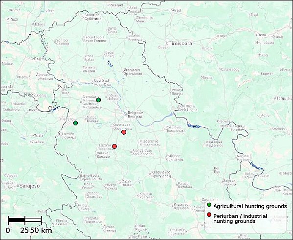
Figure 1. Map of Central Serbia illustrating the locations of the investigated hunting grounds.
Study design
The study was designed as a cross-sectional observational study with a comparative approach. The aim was to determine whether the frequency and patterns of pathomorphological changes in the organs of the European brown hare (
Study population and animal characteristics
All
Differences in sample size among hunting ground types resulted from several interconnected factors, including variations in population status, abundance, and culling plans across individual hunting grounds. These factors also affected the number of organized hunting days during the season and harvest success, which was further influenced by terrain features and the number of participants in hunting activities. Therefore, the final sample size and its distribution by hunting ground type reflect the actual management practices and field conditions during the study period.
Following harvest, in most cases only individual organs and the head were submitted, which allowed for age determination, while sex could be identified only when reproductive organs were available (Table 1). Age was estimated by measuring the mass of the eye lenses using a modified method described by Mihajlović et al. [4], where individuals were classified as young (≤1 year) or adult (>1 year), with a lens mass threshold of 280 mg, in accordance with the referenced methodology. Sex was determined based on a macroscopic examination of the submitted gonads when available. Data on body condition or carcass mass were not collected, as complete carcasses were unavailable in most cases.
Table 1. Sex and age distribution of European brown hares (
| Hunting ground type | n | Male | Female | Unknown | Juvenile | Adult | Unknown |
|---|---|---|---|---|---|---|---|
| Agricultural | 25 | 6 | 7 | 12 | 7 | 6 | 12 |
| Periurban/industrial | 49 | 15 | 16 | 18 | 16 | 16 | 17 |
| Total | 74 | 21 | 23 | 30 | 23 | 22 | 29 |
The main goal of the study was to compare the frequency and distribution of pathomorphological changes across different hunting ground types. The evaluation of how individual biological factors like sex and age affected these changes was not a primary focus. Because of limited data availability and small sample sizes in some categories, sex and age were not incorporated into multifactorial statistical models. Instead, they were recorded descriptively and considered potential confounding factors when interpreting the results.
Histopathological examination
In most cases, individual organs from harvested
Because not all organs were available in every case, and due to mechanical damage during harvesting or partial degradation of some samples, the number of histopathologically examined samples varied by organ. In total, samples from 74 livers, 73 lungs, 61 spleens, and 65 kidneys were collected and analyzed.
Upon receipt, a routine macroscopic examination of the organs was performed to assess their overall appearance and guide further histopathological analysis. Since many pathological changes in the examined organs cannot be reliably identified visually, the systematic analysis in this study was mainly based on histopathological examination. Macroscopic changes were recorded when clearly visible but were not evaluated using standardized or quantitative methods, except for splenomegaly, which was documented as a binary macroscopic finding (presence or absence) and included in descriptive and statistical analyses. Conversely, for the liver, lungs, and kidneys, all pathological changes analyzed were confirmed through histopathology, and macroscopic findings in these organs were not considered as separate variables.
Liver, lung, spleen, and kidney samples intended for histopathological analysis were fixed in 10% neutral buffered formalin for 72 h, using a fixative-to-tissue ratio of at least 10:1. After fixation, samples were processed with an automatic tissue processor KD-TS3D (Kedee, PR China), which included dehydration through increasing ethanol concentrations (70%, 96%, and 100%), clearing in xylene, and paraffin embedding. The processed samples were then embedded in paraffin blocks.
Paraffin blocks were sectioned at 3–5 μm thickness using a rotary microtome Leica RM 2235 (Leica Biosystems, USA). Sections were mounted on glass slides and dried at 56°C for 24 h in a Binder E/B28 thermostat (Binder, Germany). After deparaffinization in xylene and rehydration through decreasing alcohol concentrations (100%, 96%, and 70%), sections were stained with hematoxylin and eosin (H&E). Hematoxylin was applied for 5 minutes, followed by differentiation in acid alcohol, then counterstained with eosin for 3 minutes, with water rinses between steps. After final dehydration through increasing alcohol concentrations and treatment with xylene, the slides were mounted and coverslipped. Histological sections were examined under a BX51 light microscope (Olympus Optical, Japan) and photographed using an Olympus Color View III® digital camera (Olympus, Japan). All histological procedures followed standard, widely accepted techniques for paraffin-embedded sections, as described in typical histological methodological literature [35].
Histopathological evaluation was conducted using a blinded assessment method. All slides were examined by a single trained veterinary pathologist who was unaware of the samples’ source regarding hunting ground type, thus minimizing the risk of classification bias.
Parasitic lesions were identified based on characteristic morphological features observed during histopathological examination. No additional parasitological, coprological, or molecular confirmation methods were performed; therefore, the identification of parasitic species (e.g.,
During histopathological evaluation, all observed changes in the examined organs were systematically recorded. The presence or absence of specific histopathological lesions was documented for each analyzed organ sample, and the data were compiled into a database for further analysis of lesion frequency and distribution by organ.
Statistical analysis
Statistical analysis was conducted to compare the frequency of pathomorphological changes between agricultural and periurban/industrial hunting grounds. Analyses were performed separately for each organ and lesion type, based on predefined, biologically relevant comparisons rather than an exploratory approach involving testing many variables. The choice of statistical tests was driven by the categorical nature of the data, as each pathomorphological change was considered a binary outcome (presence/absence), making tests of independence in 2×2 contingency tables the most suitable method for this analysis.
For each lesion included in the inferential analysis, a 2 × 2 contingency table (hunting ground type × lesion presence) was created. Pearson’s chi-square test was used when all expected frequencies in the table were ≥5, whereas Fisher’s exact test was employed when at least one expected frequency was <5 [36, 37]. The threshold for statistical significance was set at p < 0.05. Although multiple statistical tests were conducted, no formal correction for multiple comparisons was applied, since all analyzed pathomorphological changes were predetermined based on pathological and biological relevance and were considered within clearly defined, organ-specific hypotheses. Using conservative corrections in this study design could increase the risk of type II errors and lead to underestimating biologically significant differences between the groups.
Pathomorphological changes that had a low overall frequency were not included in inferential testing but were instead described. This method was chosen because of limited statistical power to detect differences in rare lesions and the risk of unstable estimates in contingency tables with small sample sizes, which could lead to misleading conclusions.
To improve the understanding of findings beyond just statistical significance, odds ratios (ORs) with their 95% confidence intervals (CI) were calculated for all changes included in the inferential analysis.
Statistical analyses were conducted with IBM SPSS Statistics software, version 26.0 (IBM Corp., Armonk, NY, USA).
RESULTS
Liver
Among the examined pathomorphological changes in the liver, an association was observed between dystrophic changes and hunting ground type, with hares from periurban and industrial hunting grounds showing significantly lower odds of developing these changes compared to those from agricultural hunting grounds (OR = 0.33; 95% CI: 0.12–0.89; p = 0.026). No link with hunting ground type was found for other liver changes, including hyperemia, hepatitis, and distomatosis/cholangitis, and the estimated odds ratios were accompanied by wide confidence intervals that included the value of 1.
Macroscopically, the livers were slightly enlarged, lighter in color, and fragile in texture. The main histopathological changes were dystrophic, meaning degenerative and necrotic, including intracellular edema marked by cytoplasmic clouding and vacuolization of hepatocytes, as well as necrobiotic alterations in hepatocyte nuclei such as karyopyknosis, karyorrhexis, and karyolysis, which are typical of necrosis (Figure 2b).
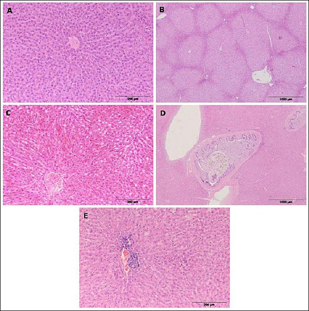
Figure 2. Representative histological sections of the liver in European brown hares. (a) Normal liver parenchyma with no visible lesions; (b) hepatic dystrophy or degeneration; (c) hepatic hyperemia or congestion; (d) hepatic distomatosis with cholangitis or pericholangitis (liver fluke infestation); (e) lymphohistiocytic hepatitis.
Along with degenerative changes, hyperemia was observed in the liver. Macroscopically, it appeared as a darker coloration of the organ and a greater amount of blood released from the cut surface. Histopathologically, it was characterized by engorgement of blood vessels (veins and arteries) and significant congestion of sinusoidal capillaries (Figure 2c).
In several individuals, inflammation of the hepatic parenchyma, known as hepatitis (Figure 2e), was observed, characterized by infiltration of mononuclear cells (lymphocytes, macrophages, and plasma cells) within the liver tissue.
Changes were also seen in the bile ducts, visible externally as wall thickening and microscopically by the presence of adult small liver fluke (
In addition to the aforementioned liver lesions, larval forms of the carnivore tapeworm (
Lungs
Analysis of pathomorphological changes in the lungs showed differences in the frequency of bronchiolitis and emphysema based on hunting ground type. These lesions were more commonly seen in hares from periurban or industrial hunting grounds, while hares from agricultural hunting grounds had lower odds of developing them, as indicated by the estimated odds ratios (bronchiolitis: OR = 0.32; 95% CI: 0.11–0.88; emphysema: OR = 0.10; 95% CI: 0.01–0.80). No link was found between hunting ground type and other tested pulmonary changes, including interstitial pneumonia, hyperemia, and hemorrhages, and the estimated odds ratios had confidence intervals that included the value of 1 (Table 3).
Table 2. Prevalence of pathomorphological lesions in the liver of European brown hares from agricultural (n = 25) and periurban/industrial (n = 49) hunting grounds, including odds ratios (OR), 95% confidence intervals (CI), and p-values.
| Lesion | Agricultural (n) | % | Periurban/ industrial (n) | % | Total (%) | OR (95% CI) | p-value |
|---|---|---|---|---|---|---|---|
| Dystrophy | 16 | 64.0 | 18 | 36.7 | 45.9 | 0.33 (0.12–0.89) | 0.026 |
| Hyperemia | 4 | 16.0 | 14 | 28.6 | 24.3 | 2.10 (0.61–7.23) | 0.233 |
| Distomatosis / cholangitis–pericholangitis | 2 | 8.0 | 10 | 20.4 | 16.2 | 2.95 (0.59–14.65) | 0.204 |
| Hepatitis | 4 | 16.0 | 6 | 12.2 | 13.5 | 0.73 (0.19–2.88) | 0.725 |
| Granulomas | 1 | 4.0 | 4 | 8.2 | 6.8 | — | — |
| Autolysis | 0 | 0.0 | 2 | 4.1 | 2.7 | — | — |
| Cysticercosis | 0 | 0.0 | 1 | 2.0 | 1.4 | — | — |
Table 3. Prevalence of pathomorphological lesions in the lungs of European brown hares from agricultural (n = 24) and periurban/industrial (n = 49) hunting grounds, including odds ratios (OR), 95% confidence intervals (CI), and p-values.
| Lesion | Agricultural (n) | % | Periurban/industrial (n) | % | Total (%) | OR (95% CI) | p-value |
|---|---|---|---|---|---|---|---|
| Bronchiolitis | 8 | 33.3 | 30 | 61.2 | 52.1 | 0.32 (0.11–0.88) | 0.025 |
| Interstitial pneumonia | 13 | 54.2 | 16 | 32.7 | 39.7 | 2.44 (0.90–6.63) | 0.078 |
| Hyperemia | 4 | 16.7 | 15 | 30.6 | 26.0 | 0.45 (0.13–1.56) | 0.202 |
| Emphysema | 1 | 4.2 | 15 | 30.6 | 21.9 | 0.10 (0.01–0.80) | 0.010 |
| Hemorrhages | 4 | 16.7 | 12 | 24.5 | 21.9 | 0.62 (0.18–2.16) | 0.448 |
| Peribronchiolitis/peribronchitis | 0 | 0.0 | 3 | 6.1 | 4.1 | — | — |
| Granulomas | 0 | 0.0 | 2 | 4.1 | 2.7 | — | — |
— indicates that statistical testing was not performed due to the low frequency of the lesion.
Bronchiolitis was the most common pathological lesion, mainly appearing as necrotizing obstructive bronchiolitis (Figure 3b), characterized by the filling of the bronchiolar lumen with desquamated necrotic epithelial cells, known as “ghost cells,” and cellular debris. In some individuals, a proliferative form of bronchiolitis was observed, marked by papillary proliferation of the bronchiolar epithelium. Peribronchiolitis and peribronchitis represented localized inflammatory processes surrounding the bronchioles and bronchi, characterized by infiltration of lymphocytes, macrophages, and plasma cells.
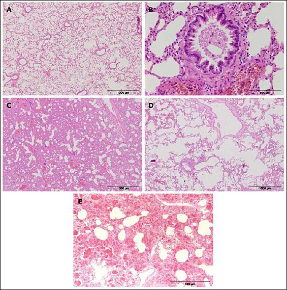
Figure 3. Representative histological sections of the lungs in European brown hares. (a) Normal pulmonary tissue; (b) necrotizing obstructive bronchiolitis; (c) interstitial pneumonia; (d) pulmonary emphysema; (e) pulmonary hemorrhage.
Interstitial pneumonia (Figure 3c) was a common finding and was characterized by thickening of the alveolar walls caused by the proliferation of type II pneumocytes, narrowing of the alveolar spaces, and increased cellularity of the interstitium, resulting in a typical “Swiss cheese” appearance of the lungs.
Among circulatory disturbances, hyperemia was the most common finding, characterized by engorgement of larger blood vessels and capillaries within the alveolar walls. Hemorrhages (Figure 3e) were evident as blood within the alveoli, bronchioles, and pulmonary interstitium.
In a smaller number of cases, changes in the form of emphysema (Figure 3d) were observed, characterized by increased airiness of the pulmonary parenchyma, with enlarged alveoli and thinning of the alveolar septa.
In some cases, granulomas were observed in the pulmonary parenchyma, composed of macrophages, epithelioid and multinucleated giant cells, lymphocytes, and a connective tissue capsule. In several individuals, cross-sections of parasites or eggs with a thick cuticle were detected in the central part of the granulomas, indicating parasitic granulomas.
Spleen
Variations in the frequency of lymphoid hyperplasia and splenomegaly in the spleen were noted depending on the type of hunting ground. These conditions were more commonly observed in hares from agricultural hunting areas, while hares from periurban or industrial hunting zones had significantly lower odds of developing them, as shown by the estimated odds ratios (hyperplasia: OR = 0.22; 95% CI: 0.07–0.70; splenomegaly: OR = 0.06; 95% CI: 0.01–0.32). Other pathological changes in the spleen did not show any association with the hunting ground type and are described narratively (Table 4).
Table 4. Prevalence of pathomorphological lesions in the spleen of European brown hares from agricultural (n = 25) and periurban/industrial (n = 36) hunting grounds, including odds ratios (OR), 95% confidence intervals (CI), and p-values.
| Lesion | Agricultural (n) | % | Periurban/industrial (n) | % | Total (%) | OR (95% CI) | p-value |
|---|---|---|---|---|---|---|---|
| Hyperplasia | 12 | 48.0 | 6 | 16.7 | 29.5 | 0.22 (0.07–0.70) | 0.008 |
| Splenomegaly | 12 | 48.0 | 2 | 5.6 | 23.0 | 0.06 (0.01–0.32) | 0.0001 |
| Hyperemia | 3 | 12.0 | 3 | 8.3 | 9.8 | — | — |
| Leukosis | 1 | 4.0 | 2 | 5.6 | 4.9 | — | — |
| Hemosiderosis | 0 | 0.0 | 2 | 5.6 | 3.3 | — | — |
— indicates that statistical testing was not performed due to the low frequency of the lesion.
Splenomegaly (Figure 4a) was a common macroscopic finding, characterized by an enlarged organ and changed tissue consistency. The spleen’s surface was smooth, while the cut surface showed dark red, moist tissue. In most cases, splenomegaly was linked to hyperplastic changes in lymphoid tissue, which histologically included lymphocyte proliferation within the white pulp, enlarged lymphoid follicles, and increased mitotic activity.
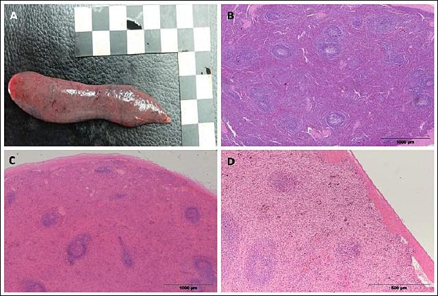
Figure 4. Representative histological sections of the spleen in European brown hares showing pathological changes. (a) Splenomegaly; (b) lymphoid hyperplasia; (c) splenic hyperemia/congestion; (d) hemosiderosis.
In some individuals, hyperplasia (Figure 4b) was accompanied by a loss of clear structural organization of the spleen, with indistinct borders between the white and red pulp, indicating significant immunoreactivity of the lymphoid tissue.
Among circulatory changes, hyperemia (Figure 4c) was observed, characterized by engorgement of blood vessels and sinusoids within the red pulp. In smaller areas of the samples, accumulation of brown pigment within macrophages was also noted, indicating splenic hemosiderosis (Figure 4d).
In rare cases, a proliferative process of lymphoid tissue with marked mitotic activity and loss of the normal cytological and histological architecture of the organ was observed, indicating a neoplastic lesion, i.e., leukosis.
Kidneys
Unlike the liver, lungs, and spleen, no clear link was found between the examined kidney changes and hunting ground type. Although variations in the frequency of tubulonephrosis, nephritis, and hyperemia were noted among the groups, the estimated odds ratios had wide confidence intervals that included the value of 1, suggesting no reliable association with hunting ground type (Table 5).
Table 5. Prevalence of pathomorphological lesions in the kidneys of European brown hares from agricultural (n = 23) and periurban/industrial (n = 42) hunting grounds, including odds ratios (OR), 95% confidence intervals (CI), and p-values.
| Lesion | Agricultural (n) | % | Periurban/industrial (n) | % | Total (%) | OR (95% CI) | p-value |
|---|---|---|---|---|---|---|---|
| Tubulonephrosis | 18 | 78.3 | 28 | 66.7 | 70.8 | 0.56 (0.16–1.96) | 0.326 |
| Nephritis (interstitial / glomerular) | 4 | 17.4 | 9 | 21.4 | 20.0 | 1.30 (0.36–4.73) | 0.758 |
| Hyperemia | 3 | 13.0 | 9 | 21.4 | 18.5 | 1.82 (0.44–7.47) | 0.515 |
| Hemorrhages | 0 | 0.0 | 1 | 2.4 | 1.5 | — | — |
— indicates that statistical testing was not performed due to the low frequency of the lesion.
Macroscopic and histopathological examination of the kidneys revealed that degenerative changes (tubulonephrosis) were the most common lesions observed (Figure 5a). The alterations affected the epithelium of the renal tubules, with microscopic evidence of vacuolar degeneration and cytoplasmic clouding. In some cases, epithelial damage was more severe, accompanied by tubulonecrosis with necrobiotic changes in the nuclei, such as karyopyknosis, karyorrhexis, and karyolysis.
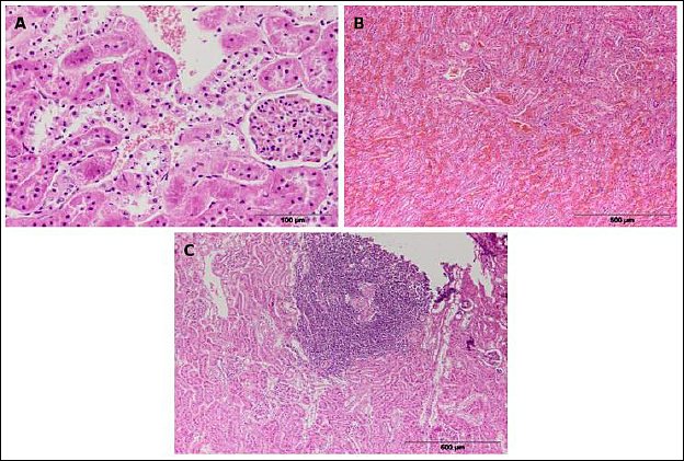
Figure 5. Representative histological sections of the kidneys in European brown hares showing pathological changes. (a) Tubulonephrosis; (b) renal hyperemia/ congestion; (c) interstitial nephritis.
Among inflammatory lesions, nephritis was the most frequently observed, most often as interstitial nephritis (Figure 5c), which was histologically characterized by focal infiltrates of lymphocytes, plasma cells, and histiocytes within the interstitium. Changes in the glomeruli appeared as proliferative or exudative glomerulonephritis.
Circulatory disturbances included hyperemia (Figure 5b), characterized by blood vessel and capillary engorgement within the renal parenchyma, while hemorrhages were rare and limited to small areas of the cortex.
DISCUSSION
Pathomorphological patterns in relation to habitat type in the European brown hare
This study examined differences in the frequency and distribution of pathomorphological changes in
Pathomorphological pattern of the European brown hare in agricultural hunting grounds
In agricultural hunting grounds, this study identified a pathomorphological pattern mainly affecting the liver and spleen. Individuals from this type of hunting area more often showed dystrophic changes in the liver, as well as reactive changes in the spleen, notably splenomegaly and lymphoid hyperplasia, compared to hares from periurban and industrial hunting grounds. The liver and spleen are interconnected organs with essential roles in metabolism, detoxification, hematopoiesis, and immune defense; therefore, changes in these organs are often considered when assessing the overall health of animal populations.
Similar types of degenerative liver changes have been observed in animal studies examining the effects of chemical substances used in agriculture, including various pesticides, where disruptions in hepatocyte structure and metabolic alterations have been reported [38–40]. Additionally, field studies on
Reactive splenic changes, including splenomegaly and hyperplasia, are generally described in the literature as adaptive responses of the organism to nonspecific stressors, without a clear etiological link to a single environmental factor [30]. However, their increased occurrence within certain habitat types may reflect differences in the overall ecological burden faced by populations, which was observed in hares from agricultural hunting grounds in this study.
Besides the pattern described, other examined histological changes in the liver and spleen did not show clear or consistent differences related to hunting ground type. Their frequency varied between agricultural and periurban/industrial habitats without forming a recognizable habitat-specific pattern. In the literature, infla-mmatory liver lesions in
In this context, although the liver and spleen are organs where reactive and adaptive changes can appear under various stressors, the pattern observed in this study suggests that certain combinations of findings may be more commonly linked to specific hunting ground types. These habitat-related “micro-patterns” should not be seen as proof of a specific cause but rather as markers indicating differences in overall ecological stress between habitat types. These findings require further validation through larger studies and by combining pathomor-phological data with information on habitat-related factors, such as pesticides typical of agricultural hunting grounds and other pollutants that might be present in different environments.
Pathomorphological pattern of the European brown hare in periurban/industrial hunting grounds
In periurban and industrial hunting grounds, the current study identified a pathomorphological pattern marked by more significant alterations in the respiratory system compared to agricultural hunting grounds. The lungs were the most affected organs, with bronchiolitis being the most prominent finding and the only lesion that clearly differed based on habitat type. Additionally, most other pulmonary lesions were generally more common in individuals from periurban and industrial hunting grounds, although without clear statistical significance, indicating a consistent but variable pattern of changes.
In this context, a study showing a higher occurrence of inflammatory lung changes in
Findings from other studies further support the idea that the respiratory system is often affected by disease changes in urban and industrial environments. In grey squirrels (
Kidneys as an organ without a habitat-specific pathomorphological pattern
Unlike certain organ systems where habitat-related patterns of changes were observed in this study, the pathomorphological lesions of the kidneys in
Similar findings have been observed in other
In this context, the even distribution of renal lesions across different hunting ground types indicates that, in this study, the kidneys of
Practical implications for hunting management and wildlife health monitoring
Based on the observed habitat-specific pathomorphological patterns, this study’s results suggest that analyzing internal organs from harvested European brown hares can be a valuable additional tool for routine population health monitoring. Along with standard indicators used in hunting management, such as population size and age structure, pathomorphological findings can offer extra insights into the health challenges faced by populations living in different habitat types.
In agricultural hunting grounds, the observed patterns of liver and spleen changes suggest that these organs may be particularly useful for assessing the health profile of populations in agroecosystems, while in periurban and industrial hunting grounds, the respiratory system may serve as a more sensitive indicator of habitat conditions. This approach allows for monitoring activities to be tailored to the dominant habitat type while still providing a comprehensive evaluation of multiple organ systems.
From a hunting management perspective, such information can help make timely adjustments to management strategies, including regulating harvest pressure and improving habitat conditions, based on the population’s health status.
Although pathomorphological findings alone do not enable the identification of specific causal factors, their systematic monitoring can serve as an early warning of potentially adverse habitat conditions, before observable declines in population size or disruptions in population structure occur. In this context, the pathomorphological findings obtained in the present study may be regarded as an additional source of information for
Given that the European brown hare is a species sensitive to land-use changes and environmental stress, the identified habitat-specific patterns of internal organ changes may help in the early detection of chronic, low-level stressors in the environment. Although this approach does not provide direct evidence of environmental degradation, it can be valuable in guiding further focused research and monitoring efforts, as well as in enhancing the long-term management and conservation of
Limitations and future perspectives
This study has certain methodological limitations that should be considered when interpreting the results. First, the assessment of environmental exposure relied on classifying hunting grounds based on land-use type, without direct measurements of potential contaminants such as pesticide residues, heavy metals, or air pollutants. As a result, it is not possible to establish direct cause-and-effect relationships between specific human activities and observed histopathological changes; instead, the results can only be interpreted within the context of habitat-related patterns.
Another limitation is the lack of screening for infectious agents. Viral, bacterial, and parasitic infections, known to cause various pathological changes in
The study was carried out during a single hunting season (2017/2018), which ensured good spatiotemporal comparability between hunting ground types and minimized the impact of climatic and weather-related confounding factors. However, this temporal limitation also prevents the assessment of seasonal and interannual variability in histopathological changes, and the results cannot be directly applied to other years or different climatic conditions without further research.
Sample size and distribution varied between hunting ground types, which may have influenced the statistical power of the analyses. Limited statistical power was especially apparent for rarely occurring histopathological lesions; therefore, findings related to low-prevalence changes should be interpreted with appropriate caution.
Histopathological changes in this study were recorded qualitatively without detailed grading of lesion severity. Although assessments followed a standardized approach, the absence of a semi-quantitative or quantitative scoring system may limit finer differentiation of lesion intensity and full reproducibility in future research.
Despite these limitations, this study offers a useful baseline for future research on histopathological patterns in
CONCLUSION
The current study showed that specific habitat-related patterns of pathomorphological changes can be observed in the European brown hare (
A major strength of this study is its cross-sectional comparative design conducted within the same geographic region and during a single hunting season, which minimized the effects of seasonal and climatic variability and allowed for more reliable comparisons between habitat types. Additionally, the simultaneous histopathological evaluation of multiple internal organs provided a comprehensive overview of the health status of the studied population, enabling the identification of organ-specific patterns rather than isolated findings. The use of routinely harvested animals also ensured that the results accurately reflect real field conditions and the actual population status, thereby enhancing the practical relevance of the findings for wildlife health monitoring and hunting management.
Although the observed differences cannot be directly linked to specific environmental factors, the results suggest that pathomorphological examination of internal organs can be a valuable complementary tool for assessing the health of European brown hare populations in habitats affected by various forms of human activity. Habitat-related patterns of lesions, especially those involving the liver, spleen, and lungs, may serve as indicative markers of the overall ecological stress experienced by populations, even without direct measurements of contaminants or other stressors.
In conclusion, this study provides evidence that systematic monitoring of the European brown hare’s health can help improve understanding of how habitat features relate to population well-being. This approach may aid wildlife management, conservation efforts, and One Health–focused environmental surveillance by allowing early detection of harmful ecological conditions and directing further focused research on potential environmental stressors.
DATA AVAILABILITY
The data generated during the study are included in the manuscript.
AUTHORS’ CONTRIBUTIONS
ZP, DB, VL, and DM: Conceptualization, study design, and methodology. NM, SS, and BS: Fieldwork, sample collection, and transport of specimens to the laboratory. DM: Pathological examinations, sample preparation, laboratory processing, organ examination, and primary interpretation of pathological findings. ZP, NM, and SS: Statistical analysis. ZP and NM: Original manuscript drafting. ZP: Supervision and coordination of the overall research process. ZP, DB, VL, DM, and NM: Interpretation of the results and formulation of recommendations. NM, ZP, and DM: Manuscript revision in response to reviewers’ comments. All authors have read and approved the final version of the manuscript.
COMPETING INTERESTS
The authors declare that they have no competing interests.
PUBLISHER’S NOTE
Veterinary World remains neutral with regard to jurisdictional claims in the published institutional affiliations.
ACKNOWLEDGMENTS
This research was supported by the Ministry of Science, Technological Development and Innovation of the Republic of Serbia (Grant No. 451-03-137/2025-03/200116).
REFERENCES
- Corbet GB. Relationships and origins of the European lagomorphs. Mammal Rev 1986;16((3-4)):105-110. [Google Scholar] | [Crossref]
- Chapman JA, Flux JEC. Rabbits, hares and pikas:Status survey and conservation action plan. Gland (Switzerland): IUCN; 1990. [Google Scholar]
- Beuković M, Beuković D, Popović Z, Đorđević N, Đorđević M. Impact of climatic factors to the percentage of young in the population of brown hare (
Lepus europaeus P.). in the Bačka district. Acta Vet 2013;63(1):111-122. [Google Scholar] | [Crossref] - Mihajlović N, StepićV V, Lavadinović V, BeukovićN N, IgnjatovićN N, Popović Z. Improving the method of lens mass preparation for age assessment in the European brown hare (
Lepus europaeus ). Acta Zool Acad Sci Hung 2023;69(4):399-412. [Google Scholar] | [Crossref] - Smith RK, Vaughan Jennings N, Harris S. A quantitative analysis of the abundance and demography of European hares (
Lepus europaeus ) in relation to habitat type, intensity of agriculture and climate. Mammal Rev 2005;35(1):1-24. [Google Scholar] | [Crossref] - Sliwinski K, Ronnenberg K, Jung K, Strauß E, Siebert U. Habitat requirements of the European brown hare (
Lepus europaeus Pallas 1778) in an intensively used agricultural region (Lower Saxony, Germany). BMC Ecol 2019;19(1):31. [Google Scholar] | [Crossref] - Panek M. Habitat factors associated with the decline in brown hare abundance in Poland in the beginning of the 21st century. Ecol Indic 2018;85:915-920. [Google Scholar] | [Crossref]
- Mayer M, Sunde P. The role of maize cultivation on European hare abundance. Agric Ecosyst Environ 2020;295:106909. [Google Scholar] | [Crossref]
- Wajdzik M, Halecki W, Kalarus K, Gąsiorek M, Pająk M. Relationship between heavy metal accumulation and morphometric parameters in European hare (
Lepus europaeus ) inhabiting various types of landscapes in southern Poland. Ecotoxicol Environ Saf 2017;145:16-23. [Google Scholar] | [Crossref] - Hornek-Gausterer R, Oberacher H, Reinstadler V, Hartmann C, Liebmann B, Lomako I. A preliminary study on the detection of potential contaminants in the European brown hare (
Lepus europaeus ) by suspect and microplastics screening. Environ Adv 2021;4:100045. [Google Scholar] | [Crossref] - Beuković D, Vukadinović M, KrstovićM M, Polovinski-Horvatović M, JajićM M, PopovićM M. The European hare (
Lepus europaeus ) as a biomonitor of lead (Pb) and cadmium (Cd) occurrence in the agro biotope of Vojvodina, Serbia. Animals (Basel) 2022;12(10):1249. [Google Scholar] | [Crossref] - Gulin J, Florijančić T, Bilandžić N, Ozimec S, Bošković I, Lončarić Z. Heavy metals (As, Cd, Hg, and Pb) in hare tissues:A survey. Poljoprivreda (Osijek) 2023;29(2):86-96. [Google Scholar] | [Crossref]
- Santilli F, Viviano A, Mori E. Dietary habits of the European brown hare:Summary of knowledge and management relapses. Ethol Ecol Evol 2024;36(1):21-40. [Google Scholar] | [Crossref]
- Edwards PJ, Fletcher MR, Berny P. Review of the factors affecting the decline of the European brown hare (
Lepus europaeus Pallas, 1778) and the use of wildlife incident data to evaluate the significance of paraquat. Agric Ecosyst Environ 2000;79((2-3)):95-103. [Google Scholar] | [Crossref] - Langhammer M, Grimm V. Mitigating bioenergy-driven biodiversity decline:A modelling approach with the European brown hare. Ecol Model 2020;416:108914. [Google Scholar] | [Crossref]
- Johann F, Arnold J. Scattered woody vegetation promotes European brown hare population. Basic Appl Ecol 2021;56:322-334. [Google Scholar] | [Crossref]
- Gryz J, Krauze-Gryz D. Why did brown hare (
Lepus europaeus ) disappear from some areas in central Poland?Diversity (Basel). 2022;14(6):465. [Google Scholar] | [Crossref] - Hakme E, Pagh S, Cutillas VM, Haydar S, Hansen MS, Larsen HL. Exposure of European hares (
Lepus europaeus ) to pesticides in Danish agricultural areas. Environ Pollut 2025;381:126597. [Google Scholar] | [Crossref] - Pagh S, Jensen LL, Pertoldi C, Larsen HL. Monitoring urban European hares (
Lepus europaeus Pallas) with citizen science and a thermal spotter. Urban Sci 2025;9(2):27. [Google Scholar] | [Crossref] - Tsokana C, Sokos C, Giannakopoulos A, Birtsas P, Valiakos G, Spyrou V. European brown hare (
Lepus europaeus ) as a source of emerging and re-emerging pathogens of public health importance:A review. Vet Med Sci 2020;6(3):550-564. [Google Scholar] | [Crossref] - Ristić Z, Ponjiger I, Matejević M, Kovačević M, Ristić N, Marković V. Effects of factors associated with the decline of brown hare abundance in the Vojvodina region (Serbia). Hystrix It J Mammal 2021;32(1):67-71. [Google Scholar] | [Crossref]
- Voigt U, Siebert U. Survival rates of pre-weaning European hares (
Lepus europaeus ) in an intensively used agricultural area. Eur J Wildl Res 2020;66((4)):67. [Google Scholar] | [Crossref] - Ilić T, Petrović T, Dimitrijević S. Parasitic infections of wild rabbits and hares. Vet Glas 2014;68((3-4)):241-250. [Google Scholar] | [Crossref]
- Marinković D, RelićD D, LakićD D, AničićD D, BeukovićD D, Davidović D. Influence of age, habitat elevation, and distance to a thermal power plant on pathomorphological findings in the European brown hare (
Lepus europaeus P.). Vet Q 2023;43(1):1-10. [Google Scholar] | [Crossref] - Lamarque F, Barrat J, Moutou F. Principal diagnoses for determining causes of mortality in the European hare (
Lepus europaeus ) found dead in France between 1986 and 1994. Gibier Faune Sauvage, Game Wildl Sci 1996;13(1):53-72. [Google Scholar] | [Crossref] - Fitzner A, Niedbalski W, Kęsy A, Rataj B, Flis M. European brown hare syndrome in Poland:Current epidemiological situation. Viruses (Basel) 2022;14(11):2423. [Google Scholar] | [Crossref]
- Faehndrich M, Klink JC, Roller M, Wohlsein P, Raue K, Strube C. Status of infectious diseases in free-ranging European brown hares (
Lepus europaeus ) found dead between 2017 and 2020 in Schleswig-Holstein, Germany. Pathogens 2023a;12((2)):239. [Google Scholar] | [Crossref] - Vizzarri F, Slamecka J, Sladecek T, Jurcik R, Ondruska L, Schultz P. Long-term monitoring of European brown hare (
Lepus europaeus ) population in the Slovak Danubian Lowland. Diversity (Basel) 2024;16(8):486. [Google Scholar] | [Crossref] - Posautz A, Loncaric I, Lundin M, Hoffmann D, Lavazza A, Kelemen Z. Health screening of free-ranging European brown hares (
Lepus europaeus ) on the German North-Sea island Pellworm. Acta Vet Scand 2015;57(1):43. [Google Scholar] | [Crossref] - Faehndrich M, Woelfing B, Klink JC, Roller M, Baumgärtner W, Wohlsein P. Pathomorphological findings and infectious diseases in selected European brown hare (
Lepus europaeus Pallas, 1778) populations from Schleswig-Holstein, Germany. Pathogens 2023b;12((11)):1317. [Google Scholar] | [Crossref] - Lekko YM, Ooi PT, Omar S, Mazlan M, Ramanoon SZ, Jasni S.
Mycobacterium tuberculosis complex in wildlife:Review of current applications of antemortem and postmortem diagnosis. Vet World 2020;13(9):1822-1836. [Google Scholar] | [Crossref] - Linšak Ž, Gobin I, Linšak DT, Broznić D. Effects of long-term lead exposure on antioxidant enzyme defense system in organs of brown hare (
Lepus europaeus Pallas) as a bioindicator of environmental pollution in Croatia. Biol Trace Elem Res 2022;200((12)):5091-5103. [Google Scholar] | [Crossref] - Can SÜ, Demirbaş Y, Erduran N, Beuković D. The European Hare (
Lepus europaeus ) as a Biomonitor of Selected Heavy Metal Pollution in the Marmara Region, Türkiye. Contemp Agric 2025;74((1-2)):35-44. [Google Scholar] | [Crossref] - Law on Game and Hunting (Zakon o divljači i lovstvu) [Serbian. Off Gaz Repub Serbia 2018;18. [Google Scholar] | [Crossref]
- Bancroft JD, Gamble M. Theory and practice of histological techniques. Elsevier; p. 2019. [Google Scholar]
- McDonald JH. Handbook of Biological Statistics. Baltimore (MD): Sparky House Publishing; p. 2014. [Google Scholar]
- Agresti A. An introduction to categorical data analysis. Hoboken (NJ): Wiley; p. 2019. [Google Scholar]
- Al-Arami AM, Al-Sanabani AS. Histopathological effects of pesticide imidacloprid insecticide on the liver in male rabbits. Ibn Al-Haitham J Pure Appl Sci 2021;34(4):1-9. [Google Scholar] | [Crossref]
- Akter L, Kobir MA, Nasrin M, Siddiqi MNH, Pervin M, Karim MR. Effects of exposure to imidacloprid-contaminated feed on the visceral organs of adult male rabbits (
Oryctolagus cuniculus ). Saudi J Biol Sci 2023;30(7):103684. [Google Scholar] | [Crossref] - Alarcan J, Sprenger H, Waizenegger J, Lichtenstein D, Luckert C, Marx-Stoelting P. Transcriptomics analysis of hepatotoxicity induced by the pesticides imazalil, thiacloprid and clothianidin alone or in binary mixtures in a 28-day study in female Wistar rats. Arch Toxicol 2021;95(3):1039-1053. [Google Scholar] | [Crossref]
- Gustafsson K, Svensson T, Uggla A. Studies on an idiopathic syndrome in the brown hare (
Lepus europaeus P.) and mountain hare (Lepus timidus L.) in Sweden, with special reference to hepatic lesions. J Vet Med A 1989;36((1-10)):631-637. [Google Scholar] | [Crossref] - Torres-Blas I, Horsler H, Paredes UM, Perkins M, Priestnall SL, Brekke P. Impact of exposure to urban air pollution on grey squirrel (
Sciurus carolinensis ) lung health. Environ Pollut 2023;326:121312. [Google Scholar] | [Crossref] - Thapa S, Bhatta R, Puri B, Bashyal R, Kunwar R, Shrestha SP. A pilot study on pulmonary anthracosis in stray dogs of Kathmandu Valley, Nepal:A potential public health threat for the future. Vet World 2024;17(3):658-665. [Google Scholar] | [Crossref]
- Vignal C, Pichavant M, Alleman LY, Djouina M, Dingreville F, Perdrix E. Effects of urban coarse particles inhalation on oxidative and inflammatory parameters in the mouse lung and colon. Part Fibre Toxicol 2017;14(1):46. [Google Scholar] | [Crossref]
- Lin CI, Tsai CH, Sun YL, Hsieh WY, Lin YC, Chen CY. Instillation of particulate matter 2.5 induced acute lung injury and attenuated the injury recovery in ACE2 knockout mice. Int J Biol Sci 2018;14(3):253-265. [Google Scholar] | [Crossref]
- Selmi S, Rtibi K, Grami D, Sebai H, Marzouki L. Malathion, an organophosphate insecticide, provokes metabolic, histopathologic and molecular disorders in liver and kidney in prepubertal male mice. Toxicol Rep 2018;5:189-195. [Google Scholar] | [Crossref]
- Dong G, Li Q, Yu C, Wang Q, Zuo D, Li X. N-acetylcysteine protects against diazinon-induced histopathological damage and apoptosis in renal tissue of rats. Toxicol Res 2024;40((2)):285-295. [Google Scholar] | [Crossref]
- Bal SS, Leishangthem GD, Singh A. Fipronil-induced kidney injury in mice and its attenuation with p-coumaric acid. J Basic Appl Zool 2025;86:34. [Google Scholar] | [Crossref]
- Sharaf S, Khan MUR, Aslam A, Rabbani M, Sharf A, Ijaz M. Toxico-pathological effects of heavy metals from industrial drainage wastewater on vital organs of small ruminants in Lahore. Environ Sci Pollut Res;2021(28):3533-3543. [Google Scholar] | [Crossref]
- Beiglböck C, Steineck T, Tataruch F, Ruf T. Environmental cadmium induces histopathological changes in kidneys of roe deer. Environ Toxicol Chem 2002;21(9):1811-1816. [Google Scholar] | [Crossref]