

ABSTRACT
Background and Aim: The coconut lorikeet (
Materials and Methods: Six clinically healthy captive
Results: A total of 1859 bacterial species were identified across all groups. Microbial composition differed markedly by age and anatomical site. Cloacal samples in both adults and juveniles were dominated by
Conclusion: This study provides the first comprehensive characterization of oral and cloacal microbiomes in captive
Keywords: age-related variation, avian microbiome, captive breeding, cloacal microbiota, coconut lorikeet, microbial diversity, nectarivore, oral microbiota.
INTRODUCTION
Coconut lorikeets (
Such dietary modifications can directly influence host-associated microorganisms. Previous studies have reported only minor differences in gut microbiota between wild and captive birds [4]. However, microbiome composition and diversity are strongly influenced by anatomical site, diet, and age [5, 6]. Adaptations in the digestive system of lorikeets include a less muscular gizzard and a shorter intestine compared with granivorous and frugivorous parrots [7]. Their ability to digest sucrose is determined by gut sucrase activity [8, 9]. Under high sugar concentrations, rainbow lorikeets preferentially consume hexose over sucrose to optimize digestion [9, 10]. This dietary selectivity may influence foraging behavior, as birds select flowers with optimal sugar composition. Consequently, the abundance and distribution of flowering plants play a crucial role in survival and reproduction, thereby influencing feeding patterns [3].
Microorganisms contribute significantly to host physiology by enhancing growth, stress tolerance, and feed efficiency [11, 12]. In many species, the oral microbiome serves as an indicator of oral and systemic health [13–15], while the gut microbiome is closely linked to dietary habits [16].
Although previous studies have explored avian microbiomes, there is a lack of integrated analysis focusing on both oral and cloacal microbiota in nectarivorous parrots under controlled captive conditions. Existing literature has primarily emphasized gut microbiota, often overlooking the oral cavity, which plays a critical role in early microbial colonization and dietary interactions. Furthermore, the combined influence of age and anatomical site on microbial diversity in
Therefore, this study aimed to identify, characterize, and compare the oral and cloacal microbiomes of adult and juvenile
MATERIALS AND METHODS
Ethical approval
All experimental procedures involving animals were reviewed and approved by the Animal Ethics Committee of the Indonesian Agency for Research and Innovation (BRIN), Bogor, Indonesia (Approval Number: 020/KE.02/SK/8/2022). The study was conducted in full compliance with national animal welfare regulations and internationally accepted ethical guidelines, including the ARRIVE guidelines 2.0 and the standards established by the World Organization for Animal Health (WOAH).
The study involved non-invasive sampling procedures using oral and cloacal swabs, which did not cause pain, injury, or long-term distress to the animals. Handling of
All birds were clinically healthy and sourced from a monitored captive population. Prior to inclusion, animals were assessed by a veterinarian to confirm the absence of disease or abnormal physiological conditions. Throughout the study period, birds were maintained under controlled environmental conditions and monitored daily for behavioral changes, physical abnormalities, and signs of distress. Any indication of compromised welfare would have resulted in immediate veterinary intervention; however, no such events occurred.
Housing, feeding, and environmental enrichment were provided in accordance with best practices for captive avian management. The study design adhered to the principles of the 3Rs (Replacement, Reduction, and Refinement), using the minimum number of animals required to achieve scientific objectives while ensuring high standards of care and minimizing animal use.
No endangered or protected individuals were harmed, and the study did not involve wild capture or invasive manipulation. All procedures were conducted in a manner that ensured the highest level of ethical responsibility, animal welfare, and scientific integrity.
Study period and location
The study was conducted from July to October 2024 at a controlled research facility under the Indonesian Agency for Research and Innovation (BRIN), Bogor, Indonesia. The experimental and sampling activities were performed during a continuous 14-day observation period under standardized environmental conditions.
Study design
This study employed a controlled experimental design involving six clinically healthy

Figure 1. Schematic diagram of sampling, DNA extraction, and pooling samples
Bird care and housing
A total of six
Feed intake for each bird was recorded daily over 14 days. The quantities of feed offered and remaining were weighed, and intake was calculated as the difference between these values. All birds were sourced from a monitored population and showed no clinical signs of disease. Additional diagnostic screening was deemed unnecessary by the attending veterinarian. Health monitoring included daily observation of morphology, behavior, and clinical signs of stress or disease. All animals remained under continuous veterinary supervision throughout the study.
Sampling and handling
Six
Each swab was placed into a sterile tube containing 10% glycerol as a cryoprotectant and stored at −80°C for long-term preservation until DNA extraction.
Sex determination
Sex determination was performed using polymerase chain reaction (PCR) with universal primers 2550F (5’-GTTACTGATTCGTCTACGAGA-3’) and 2718R (5’-ATTGAAATGATCCAGTGCTTG-3’). These primers target the CHD gene, which exists in two forms: CHD-Z (Z chromosome) and CHD-W (W chromosome). Breast feather samples (plumae) were used for DNA extraction.
DNA extraction
DNA extraction was performed individually for each sample before pooling. Extraction was carried out using the QIAamp Fast DNA Stool Mini Kit (Qiagen, Hilden, Germany) according to the manufacturer’s protocol with modifications, including treatment with lysozyme (25 mg/mL) (Calbiochem, Millipore, USA and Canada), RBC lysis buffer, and RNase (10 µL/mL) (Geneaid Biotech Ltd., Taipei, Taiwan).
This process yielded three JO, three JC, three AO, and three AC DNA extracts. DNA concentration and purity were measured using a nanophotometer (Implen, Munich, Germany). After quality assessment, equal amounts of DNA from individuals within each group were pooled, resulting in four composite samples: JO, JC, AO, and AC.
Bioinformatic pipeline
Sequencing libraries were prepared following standard protocols. Sequencing was performed using ONT with the PromethION platform. Raw signal data were acquired using MinKNOW software version 24.02.16 in FAST5 format. Basecalling was conducted using Dorado version 7.3.11 [18], generating FASTQ files under a high-accuracy model (Table 1).
Table 1. Summary of raw and filtered sequencing data.
| Group | n | Raw data (reads) | Mean read length (bp) | Filtered data (reads) | Mean read length (bp) |
|---|---|---|---|---|---|
| AO | 3 | 102,700.0 | 1,589.7 | 93,712.0 | 1,610.0 |
| JO | 3 | 102,700.0 | 1,593.8 | 94,773.0 | 1,609.5 |
| AC | 3 | 105,846.0 | 1,576.9 | 94,803.0 | 1,610.9 |
| JC | 3 | 104,204.0 | 1,563.4 | 91,525.0 | 1,625.0 |
Quality control was performed using NanoPlot [19, 20] to evaluate read length and quality distribution. Reads were filtered using NanoFilt version 1.8.0 (https://github.com/wdecoster/nanoflt), with a minimum read length of 1000 bp and minimum quality score threshold. High-quality reads were taxonomically classified using Centrifuge against the SILVA 16S rRNA reference database with a confidence threshold of 0.7 [21].
Taxonomic abundance was visualized using Pavian, and radial community structures were generated using KronaTools. Downstream analyses included alpha diversity (Observed operational taxonomic unit (OTUs), abundance-based coverage estimator (ACE), Simpson, and Fisher) and beta diversity (Venn diagram and principal coordinate analysis (PCoA)) using R software version 4.2.3 (https://www.R- project.org/). No inferential statistical tests were performed; all analyses were descriptive.
PCR product visualization
PCR products were analyzed using gel electrophoresis on 1% TBE agarose. Migration patterns of samples (lanes 1–4) were compared with a 1 Kb DNA ladder (lane M) to determine fragment size. The non-template control confirmed the absence of contamination (Figure 2).

Figure 2. Polymerase chain reaction product (amplification of gDNA with primer 16S rRNA 27F–1492R).
Statistical analysis
This study used a bioinformatics-based metagenomic approach. Sequencing data were processed through ONT pipelines including MinKNOW, Dorado, NanoPlot, and NanoFilt version 1.8.0 (https://github.com/ wdecoster/nanoflt). Taxonomic classification was performed using Centrifuge with the SILVA 16S rRNA database, followed by downstream analysis using Pavian https://github.com/fbreitwieser/pavian), KronaTools (https:// github.com/marbl/Krona), and R software version 4.2.3 (https://www.R- project.org/).
Alpha diversity (Observed OTUs, ACE, Simpson, and Fisher) and beta diversity (Venn diagram and PCoA) were calculated using R Studio (version 4.2.3). No inferential statistical analysis was conducted, and all results are presented descriptively.
RESULTS
Biological characteristics of experimental birds
The biological data of the experimental subjects are presented in Table 2. The data include sex, age, body weight, and rearing period for both juvenile and adult groups.
Table 2. Biological data of juvenile and adult
| Group | ID | Sex | Age | Weight | Rearing period |
|---|---|---|---|---|---|
| Juvenile | J1 | Male | 2 months | 115 g | 2 months |
| J2 | Female | 3 months | 117 g | 3 months | |
| J3 | Male | 2 months | 113 g | 2 months | |
| Adult | A1 | Male | Adult | 140 g | ± 3 years |
| A2 | Female | Adult | 130 g | ± 3 years | |
| A3 | Female | Adult | 130 g | ± 3 years |
* Born in research facility,
** Born in commercial facility
Feed intake patterns
The average daily feed intake data for juvenile and adult
Table 3. Daily feed intake of juvenile and adult
| Feed | Juvenile (X ± sd) | Adult (X ± sd) |
|---|---|---|
| Apple | 0.34 ± 0.16 | 2.78 ± 0.44 |
| Mango | 0.71 ± 0.32 | 1.87 ± 0.40 |
| Papaya | 2.04 ± 1.29 | 2.25 ± 0.36 |
| Banana | 1.11 ± 0.71 | 2.84 ± 0.76 |
| Guava | 1.78 ± 0.27 | 1.72 ± 0.33 |
| Fresh corn | 3.78 ± 0.89 | 5.95 ± 2.83 |
| Sunflower seed | 0 | 2.97 ± 0.51 |
| Cooked edamame | 1.18 ± 0.81 | 4.97 ± 0.63 |
| Bean sprouts | 0.14 ± 0.01 | 1.65 ± 0.40 |
| Cucumber | 2.05 ± 1.89 | 3.98 ± 1.86 |
| Long bean | 0 | 1.83 ± 0.57 |
| Baby biscuit in nectar solution | 47.12 ± 10.94 | 70.34 ± 7.56 |
Nutritional composition of feed
The nutritional content of
Table 4. Nutritional composition of
| Nutrient | Unit | Apple* | Mango* | Papaya* | Banana* | Guava* | Fresh corn* | Sunflower seed* | Cooked edamame* | Bean sprouts* | Cucumber* | Long bean* | Baby biscuit in nectar |
|---|---|---|---|---|---|---|---|---|---|---|---|---|---|
| Macronutrients | |||||||||||||
| Water | gram | 86.00 | 83.46 | 89.80 | 75.30 | 84.25 | 76.05 | 1.20 | 72.77 | 90.40 | 95.23 | 87.85 | 74.28 |
| Total fat | gram | 0.20 | 0.38 | 0.14 | 0.26 | 0.95 | 1.38 | 50.00 | 5.20 | 0.19 | 0.19 | 0.44 | 0.57 |
| Protein | gram | 0.26 | 0.85 | 0.60 | 0.78 | 2.55 | 3.24 | 19.53 | 12.00 | 3.08 | 0.58 | 2.80 | 0.40 |
| Total carbohydrate | gram | 13.81 | 15.50 | 10.98 | 22.61 | 14.32 | 18.62 | 24.22 | 8.91 | 5.96 | 3.65 | 8.35 | 24.66 |
| Calories | kcal | 0.05 | 0.06 | 0.04 | 0.10 | 0.07 | 0.09 | 0.58 | 0.14 | 0.03 | 0.02 | 0.05 | 105.33 |
| Dietary fibers | gram | 2.40 | 1.58 | 0.80 | 1.74 | 5.40 | 2.00 | 10.94 | 5.20 | 1.83 | 0.58 | 4.00 | – |
| Sugar | gram | 10.39 | 13.94 | 9.80 | 15.82 | 8.92 | 6.28 | 2.73 | 2.18 | 4.13 | 1.73 | 1.88 | – |
| Ash | gram | 0.30 | 0.36 | 0.70 | 0.70 | 0.65 | 0.62 | 5.60 | 1.21 | 0.44 | 0.38 | 0.58 | 0.10 |
| Micronutrients - Vitamins | |||||||||||||
| Folate | mcg | 3.00 | 43.00 | 55.00 | 23.60 | 49.00 | 42.00 | 237.00 | 311.00 | 61.00 | 7.00 | 62.00 | 122.64 |
| Niacin (Vit B3) | mg | 0.09 | 0.67 | 0.34 | 0.66 | 1.08 | 1.77 | 7.04 | 0.96 | 0.75 | 0.10 | 0.41 | 0.00 |
| Riboflavin (Vit B2) | mg | 0.03 | 0.10 | 0.07 | 0.00 | 0.04 | 0.06 | 0.25 | 0.16 | 0.12 | 0.03 | 0.11 | 0.57 |
| Thiamin (Vit B1) | mg | 0.02 | 0.03 | 0.05 | 0.06 | 0.07 | 0.16 | 0.11 | 0.20 | 0.08 | 0.03 | 0.11 | 0.00 |
| Vitamin A | IU | 208.39 | 180.00 | 156.70 | 3.33 | 103.32 | 29.97 | 0.00 | 866.66 | 3.33 | 16.65 | 143.19 | 235.34 |
| Vitamin B6 | mg | 0.04 | 0.12 | 0.15 | 0.21 | 0.11 | 0.09 | 0.80 | 0.10 | 0.09 | 0.04 | 0.02 | 0.49 |
| Vitamin C | mg | 4.60 | 36.42 | 108.00 | 12.26 | 228.30 | 6.83 | 1.41 | 6.10 | 13.17 | 2.88 | 18.80 | 4.12 |
| Vitamin E | mg | 0.18 | 0.90 | 5.30 | 0.20 | 0.73 | 0.07 | 26.10 | 0.68 | 0.10 | 0.04 | 0.00 | 6.09 |
| Vitamin K | mcg | 2.20 | 4.18 | 2.90 | 0.09 | 2.60 | 0.28 | 2.73 | 26.70 | 33.08 | 16.35 | 41.60 | 5.09 |
| Micronutrients - Minerals | |||||||||||||
| Calcium, Ca | mg | 6.00 | 10.91 | 24.00 | 5.00 | 18.00 | 2.00 | 70.00 | 63.00 | 13.00 | 16.00 | 50.00 | 201.27 |
| Copper, Cu | mg | 0.03 | 0.11 | 0.14 | 0.10 | 0.23 | 0.06 | 1.83 | 0.35 | 0.16 | 0.04 | 0.05 | 0.00 |
| Iron, Fe | mg | 0.12 | 0.16 | 0.66 | 0.00 | 0.26 | 0.52 | 3.80 | 2.27 | 0.91 | 0.29 | 0.47 | 6.09 |
| Magnesium, Mg | mg | 5.00 | 10.00 | 33.00 | 28.00 | 22.00 | 37.00 | 129.00 | 64.00 | 21.00 | 13.00 | 44.00 | 28.24 |
| Manganese, Mn | mg | 0.03 | 0.06 | 0.03 | 0.26 | 0.15 | 0.16 | 2.11 | 1.02 | 0.19 | 0.08 | 0.21 | 0.33 |
| Phosphorus, P | mg | 11.00 | 14.00 | 33.00 | 22.00 | 40.00 | 89.00 | 1155.00 | 169.00 | 54.00 | 24.00 | 59.00 | 78.63 |
| Potassium, K | mg | 107.00 | 168.00 | 257.00 | 326.00 | 417.00 | 270.00 | 850.00 | 436.00 | 149.00 | 147.00 | 240.00 | 0.00 |
| Selenium, Se | mcg | 0.00 | 0.60 | 1.50 | 0.00 | 0.60 | 0.60 | 79.30 | 0.80 | 0.60 | 0.31 | 1.50 | 0.00 |
| Sodium, Na | mg | 1.00 | 1.00 | 3.00 | 0.00 | 2.00 | 15.00 | 3.00 | 6.00 | 6.00 | 2.00 | 4.00 | 0.00 |
| Zinc, Zn | mg | 0.04 | 0.09 | 0.09 | 0.16 | 0.23 | 0.46 | 5.29 | 1.37 | 0.41 | 0.19 | 0.37 | 2.45 |
Overall microbial diversity
The gut microbiota of
Alpha diversity analysis
Alpha diversity of the gut microbiome in
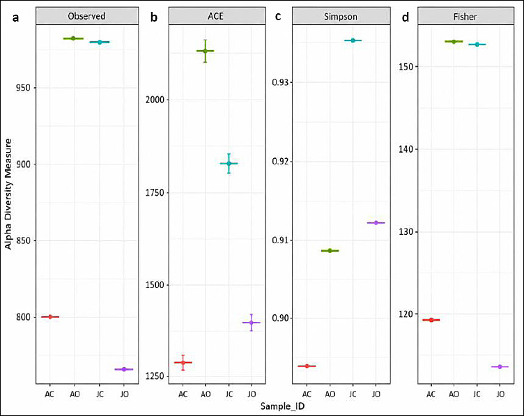
Figure 3. General information of alpha diversity analysis of bacterial gut microbiome of
Observed OTUs showed that JC and AO had similar values (~980) and the highest richness, whereas AC and JO exhibited lower values (~800). The ACE index indicated higher richness in JC (1829) and AO (2131), while AC and JO showed lower richness (1283 and 1358, respectively). The Simpson index demonstrated that JC had the highest diversity value, whereas JO and AO had similar values (0.91), and AC showed the lowest diversity. The Fisher index indicated higher diversity in AO (153.00) and JC (1522.70), with AC and JO showing lower values (119.35 and 113.21, respectively).
Beta diversity analysis
Beta diversity of microbiota samples in

Figure 4. Venn diagram showing relationships between samples at the species level: (a) all samples; (b) JC vs JO; (c) AC vs AO; (d) AC vs JC; (e) AO vs JO. AC: Adult cloaca; AO: Adult oral; JC: Juvenile cloaca; JO: Juvenile oral.
JC and JO shared 246 core OTUs, whereas AC and AO shared 220 OTUs. JC exhibited higher unique OTUs than JO (734 vs 516), while AC had fewer unique OTUs than AO (580 vs 762). Furthermore, AC and JC shared 493 OTUs, and AO and JO shared 365 OTUs. AC had fewer unique OTUs than JC (307 vs 487), whereas AO had more unique OTUs than JO (617 vs 397).
Microbial community composition
The relative abundance of bacterial communities was analyzed at genus and species levels (Figures 5a and b). Microbial composition differed across AO, AC, JO, and JC groups.

Figure 5. (a) Relative abundance of bacterial communities at the genus level (top 10 genera). (b) Relative abundance of bacterial communities at the species level (top 10 species).
At the genus level,
JC exhibited a more complex microbial structure, dominated by
At the species level, AC was dominated by
JC was dominated by
Heatmap visualization of microbial abundance
The top 50 OTUs were visualized using a heatmap (Figure 6). Color intensity reflects microbial abundance, with lighter colors indicating higher abundance. Distinct microbial patterns were observed across age groups and sampling sites.

Figure 6. Heatmap of relative abundance of
Targeted microbial populations
The specific pathogenic bacteria taxa identified across all samples, along with their associated references are summarized in Table 5 [22, 24–30]. This table provides a detailed overview of these potential pathogens, highlighting their relevance to the health monitoring of captive birds.
Table 5. Population of targeted gut microbes
| Genus | Adult cloaca | Adult oral | Juvenile cloaca | Juvenile oral | Reference |
|---|---|---|---|---|---|
| 59 | 1 | 226 | 1 | [22–24] | |
| 19 | 0 | 1692 | 0 | [22] | |
| 1 | 7 | 1042 | 3 | [25] | |
| 27 | 0 | 29 | 0 | [25] | |
| 8 | 282 | 12 | 40 | [22, 24, 26] | |
| 10539 | 33 | 5625 | 41 | [27] | |
| 1 | 0 | 0 | 0 | [28] | |
| 55 | 9 | 519 | 87 | [22, 29] | |
| 23 | 0 | 9 | 2 | [22, 30] |
PCoA analysis
PCoA analysis demonstrated distinct clustering patterns among sample groups (Figure 7). The first axis explained 79.64% of variance, while the second and third axes explained 11.21% and 9.16%, respectively. These results indicate clear separation among microbial communities, primarily driven by sampling site and age.

Figure 7. PCoA plots based on Bray–Curtis dissimilarity.
DISCUSSION
Age- and site-related variation in microbial diversity
Based on the present results, the oral microbiota of adult
Conversely, the oral microbiota of juvenile
The cloacal microbiota of adult (AC) and juvenile (JC)
Microbial stability and developmental effects in juveniles
The present results also indicated that the gut microbiome of juvenile birds remains strongly influenced by environmental factors and feeding behavior. The high alpha diversity observed in juvenile cloacal samples of
Gastrointestinal tracts of juvenile birds are initially colonized by numerous transient bacterial species, which gradually develop into a more stable adult-like community [36]. In addition to bacteria, other studies have shown that juvenile birds harbor more transient viral species, probably acquired early in life, whereas adults exhibit a more stable viral community. These changes in microbial diversity reflect the influence of age on both bacterial and viral communities within avian hosts [37]. Meanwhile, the high alpha diversity observed in the oral microbiome of adult birds may indicate more varied feeding behavior in adults than in juveniles [38].
Ecological relevance of Rosenbergiella in nectar-feeding birds
Influence of habitat, diet, and anatomical site on microbiome composition
Microbiome diversity is strongly influenced by environmental factors, including habitat and lifestyle. Birds with broader habitat ranges and more diverse diets typically have more diverse microbiomes [44]. The diversity of microbiota in birds differs substantially between the digestive tract and oral cavity because of differences in pH, enzymes, and organic substrates available in each habitat [45]. Dominant bacteria in the oral cavity of vertebrates often belong to Firmicutes and Bacteroidetes, whereas Proteobacteria are usually more common in the cloaca or digestive tract. These phyla are frequently found in avian gut ecosystems and play important roles in digestion and immune function. Differences in the dominance of these genera or species can be influenced by diet, lifestyle, and environmental conditions [46, 47].
Potential probiotic relevance of Weissella and other commensal bacteria
The presence of
The gut microbiome is crucial for avian health because it influences disease resistance and overall well-being. In chickens, modulation of the gut microbiota has been shown to alleviate infections, highlighting the potential role of microbiome management in disease control [22]. Microbiomes also contribute to ecological interactions, including pathogen transmission. Migratory birds can act as reservoirs for pathogens, posing risks to other species and humans [50, 51]. Similar to mammalian systems, the gut microbiota supports immune system function and nutrient absorption. It contributes to general physiological health and assists in the processing of pollutants [52]. The microbiome also protects against pathogens, thereby supporting disease resistance. However, wild birds can harbor antibiotic-resistant bacteria, creating a risk of disease transmission and resistance spread [53].
Pathogenic bacterial taxa and implications for captive health monitoring
The gut microbiome of wild birds is highly diverse and dynamic, playing a crucial role in digestion, immune function, and overall health. Multiple factors, including diet, age, environmental conditions, antibiotic use, and pathogen exposure, can shape the composition of avian gut microbiota [54]. However, among the many microorganisms present in the intestine, some may act as enteropathogens and potentially cause gastrointestinal infections and other health issues [55]. These pathogenic bacteria include
Among these taxa,
Meanwhile, the prevalence of
Identification of
Furthermore, dietary changes that cause loss of host flexibility and metabolic shifts are frequently observed in captive breeding programs [58]. Captive birds can also spread antibiotic-resistant bacteria and zoonotic pathogens; therefore, mapping the gut microbiome of birds intended for reintroduction has become a useful tool for wildlife conservation and the One Health concept [59].
Antibiotic resistance and One Health relevance
Several recent studies have demonstrated that captivity and environmental transition can substantially influence the prevalence and distribution of antibiotic resistance in wildlife. One study identified that rehabilitation and environmental changes in migratory birds were associated with increased antibiotic resistance, including the emergence of multidrug resistance in
Young birds may have different diets and foraging behaviors than adults, which can increase their exposure to contaminated feed sources. The cloacal microbiota of juvenile birds is often more diverse and less stable than that of adults. This diversity may include a higher prevalence of pathogenic bacteria, such as
Commensal bacteria are integral components of the gut microbiome in wild birds and contribute to physiological functions such as digestion and immune modulation. Among these,
PCoA-based interpretation of community structure
The separation along PCoA1 suggests that this axis represents a key ecological gradient influencing microbial community composition. Clustering of samples within the same group indicates that community structures are more similar within groups than between groups, suggesting that specific environmental or biological factors shape microbial composition. PCoA results in migratory birds indicated that the microbial structure of migratory birds was more heterogeneous than that of environmental samples [62]. Overall, this pattern suggests that the main factor distinguishing the groups plays a significant role in shaping community structure. Samples within the same group tended to cluster together because of shared characteristics, whereas clear separation between groups reflected distinct compositional differences.
There was clear separation of sample points, suggesting that the community composition of AC, AO, JC, and JO differed distinctly. Clustering of points within the same group indicated compositional similarity, whereas larger distances between groups suggested substantial differences. Separation along PCoA1 indicates that this axis represents the most influential factor differentiating the groups, which may include environmental conditions, species interactions, or external influences. Gut microbes can influence phenotypic traits of animals, whereas metabolite concentrations in the gut regulate microbial functions [43, 63]. In this study, dietary changes through pellet feeding and age directly affected the intestinal microbial community and metabolites in Alexandrine parrots [43].
CONCLUSION
This study provides the first comparative characterization of oral and cloacal microbiomes in captive adult and juvenile
The dominance of nectar-associated bacteria, especially
A key strength of this study is the use of full-length 16S rRNA sequencing, which improved taxonomic resolution and enabled detailed comparison of microbial communities across age groups and anatomical sites. The controlled housing and standardized feeding conditions also reduced environmental variation and strengthened interpretation of age- and site-associated microbial patterns.
However, this study was limited by the small number of birds, pooled sample design, and descriptive analytical approach without inferential statistical testing. In addition, 16S rRNA sequencing cannot differentiate viable from non-viable bacteria or distinguish commensal strains from pathogenic strains carrying virulence or antimicrobial resistance genes.
Future studies should include larger sample sizes, individual-level sequencing, longitudinal sampling, functional metagenomics, antimicrobial resistance profiling, and culture-based validation. Comparative studies between wild and captive
Overall, this study establishes baseline microbiome data for captive
DATA AVAILABILITY
All data generated or analyzed during this study are included in this published manuscript. Additional information, clarifications, or supplementary data can be obtained from the corresponding author upon reasonable request.
AUTHORS’ CONTRIBUTIONS
RR, SNP, SP, AF, RTP, WH, and LS: Study design. RR, KAS, APS, SM, and WW: Sample collection. RR, KAS, SP, APS, and SM: Laboratory analysis. SS, AF, and RRI: Methodology development. KAS, SS, and LS: Data validation. SNP, RRI, RTP, WH, and LS: Supervision. All authors contributed to the writing of the manuscript and approved the final version.
COMPETING INTERESTS
The authors declare that they have no competing interests.
PUBLISHER’S NOTE
Veterinary World remains neutral with regard to jurisdictional claims in the published institutional affiliations.
ACKNOWLEDGMENTS
The authors sincerely thank the National Research and Innovation Agency of Indonesia (BRIN) for financial support through the Rumah Program of the Research Organization for Life Sciences and Environment (Contract No. 1/III.5/HK/2024). The authors also thank the staff of the Department of Biology, Universitas Indonesia, for their valuable contributions during the preparation of this manuscript.
REFERENCES
- Prijono SN, Rachmatika R. Effect of sweetness level and amino acid composition of palm sugar on feed intake of
Trichoglossus haematodus in captivity. Biosaintifika 2020;12(2):254-261. [Google Scholar] - Frankel TL, Avram D. Protein requirements of rainbow lorikeets,
Trichoglossus haematodus . Aust J Zool 2001;49(4):425-443. [Google Scholar] - Davis A, Major RE, Taylor CE. The association between nectar availability and nectarivore density in urban and natural environments. Urban Ecosyst 2015;18(2):503-515. [Google Scholar]
- Garcia-Mazcorro JF, Alanis-Lopez C, Marroquin-Cardona AG, Kawas JR. Composition and potential function of fecal bacterial microbiota from six bird species. Birds 2021;2(1):42-59. [Google Scholar]
- Haberecht S, Bajagai YS, Moore RJ, Van TTH, Stanley D. Poultry feeds carry diverse microbial communities that influence chicken intestinal microbiota colonization and maturation. AMB Express 2020;10(1):143. [Google Scholar]
- Zou A, Nadeau K, Xiong X, Wang PW, Copeland JK, Lee JY. Systematic profiling of the chicken gut microbiome reveals dietary supplementation with antibiotics alters expression of multiple microbial pathways with minimal impact on community structure. Microbiome 2022;10(1):127. [Google Scholar]
- Schweizer M, Güntert M, Seehausen O, Leuenberger C, Hertwig ST. Parallel adaptations to nectarivory in parrots, key innovations and the diversification of the Loriinae. Ecol Evol 2014;4(14):2867-2883. [Google Scholar]
- McWhorter TJ, Rader JA, Schondube JE, Nicolson SW, Pinshow B, Fleming PA. Sucrose digestion capacity in birds shows convergent coevolution with nectar composition across continents. iScience 2021;24(7):102717. [Google Scholar]
- Napier KR, McWhorter TJ, Nicolson SW, Fleming PA. Sugar preferences of avian nectarivores are correlated with intestinal sucrase activity. Physiol Biochem Zool 2013;86(5):499-514. [Google Scholar]
- Nicolson SW, Fleming PA. Drinking problems on a 'simple'diet: physiological convergence in nectar-feeding birds. J Exp Biol 2014;217(7):1015-1023. [Google Scholar]
- Diaz Carrasco JM, Casanova NA, Fernández Miyakawa ME. Microbiota, gut health and chicken productivity: What is the connection?. Microorganisms 2019;7(10):374. [Google Scholar]
- Bajagai YS, Van TTH, Joat N, Chousalkar K, Moore RJ, Stanley D. Layer chicken microbiota: A comprehensive analysis of spatial and temporal dynamics across all major gut sections. J Anim Sci Biotechnol 2024;15(1):20. [Google Scholar]
- Aghili S, Rahimi H, Hakim LK, Karami S, Soufdoost RS, Oskouei AB. Interactions between oral microbiota and cancers in the aging community: A narrative review. Cancer Control 2024;31:10732748241270553. [Google Scholar]
- Xiao J, Fiscella KA, Gill SR. Oral microbiome: Possible harbinger for children's health. Int J Oral Sci 2020;12(1):12. [Google Scholar]
- Lee YH, Chung SW, Auh QS, Hong SJ, Lee YA, Jung J. Progress in oral microbiome related to oral and systemic diseases: An update. Diagnostics 2021;11(7):1283. [Google Scholar]
- Catassi G, Aloi M, Giorgio V, Gasbarrini A, Cammarota G, Ianiro G. The role of diet and nutritional interventions for the infant gut microbiome. Nutrients 2024;16(3):400. [Google Scholar]
- Bahram M, Anslan S, Hildebrand F, Bork P, Tedersoo L. Newly designed 16S rRNA metabarcoding primers amplify diverse and novel archaeal taxa from the environment. Environ Microbiol Rep 2019;11(4):487-494. [Google Scholar]
- Wick RR, Judd LM, Holt KE. Performance of neural network basecalling tools for Oxford Nanopore sequencing. [Google Scholar]
- De Coster W, D'Hert S, Schultz DT, Cruts M, Van Broeckhoven C. NanoPack: Visualizing and processing long-read sequencing data. Bioinformatics 2018;34(15):2666-2669. [Google Scholar]
- Nygaard AB, Tunsjø HS, Meisal R, Charnock C. A preliminary study on the potential of Nanopore MinION and Illumina MiSeq 16S rRNA gene sequencing to characterize building-dust microbiomes. Sci Rep 2020;10(1):3209. [Google Scholar]
- Kim D, Song L, Breitwieser FP, Salzberg SL. Centrifuge: Rapid and sensitive classification of metagenomic sequences. Genome Res 2016;26(12):1721-1729. [Google Scholar]
- Zhu J, Ding J, Yang K, Zhou H, Yang W, Qin C. Microbiome and microbial pure culture study reveal commensal microorganisms alleviate Salmonella enterica serovar pullorum infection in chickens. Microorganisms 2024;12(9):1743. [Google Scholar]
- He Y, Zhang M, Dai C, Yu L. Comparison of the gut microbial communities of domestic and wild mallards (Anas platyrhynchos) based on high-throughput sequencing technology. Animals 2023;13(18):2956. [Google Scholar]
- Cockerham S, Lee B, Orben RA, Suryan RM, Torres LG, Warzybok P. Microbial ecology of the Western Gull (Larus occidentalis). Microb Ecol 2019;78(3):665-676. [Google Scholar]
- Minich D, Madden C, Navarro MA, Glowacki L, French-Kim K, Chan W. Gut microbiota and age shape susceptibility to clostridial enteritis in lorikeets under human care. Anim Microbiome 2022;4(1):7. [Google Scholar]
- Chrobak-Chmiel D, Kwiecień E, Golke A, Dolka B, Adamczyk K, Biegańska MJ. Pigeons as carriers of clinically relevant multidrug-resistant pathogens—A clinical case report and literature review. Front Vet Sci 2021;8:664226. [Google Scholar]
- Wyres KL, Holt KE. Klebsiella pneumoniae population genomics and antimicrobial-resistant clones. Trends Microbiol 2016;24(12):944-956. [Google Scholar]
- Pak Vet J 2024. [Google Scholar]
- Fang S, Qin T, Yu T, Zhang G. Improvement of the gut microbiota in vivo by a short-chain fatty acids-producing strain Lactococcus garvieae CF11. Processes 2022;10(3):604. [Google Scholar]
- Freitas ADAR, Faria AR, Mendes LT, Merquior VLC, Neves DM, Pires JR. The gut microbiota of wild birds undergoing rehabilitation as a reservoir of multidrug-resistant enterococci in a metropolitan area in Brazil. Braz J Microbiol 2024;55(4):3849-3861. [Google Scholar]
- Koh YC, Kuo LH, Tung YC, Weerawatanakorn M, Pan MH. Identification of indicative gut microbial guilds in a natural aging mouse model. ACS Omega 2023;8(39):36569-36580. [Google Scholar]
- De La Cuesta-Zuluaga J, Kelley ST, Chen Y, Escobar JS, Mueller NT, Ley RE. Age- and sex-dependent patterns of gut microbial diversity in human adults. mSystems 2019;4(4):e00261-19. [Google Scholar]
- Zhou L, Huo X, Liu B, Wu H, Feng J. Comparative analysis of the gut microbial communities of the Eurasian kestrel (Falco tinnunculus) at different developmental stages. Front Microbiol 2020;11:592539. [Google Scholar]
- Maraci Ö, Antonatou-Papaioannou A, Jünemann S, Engel K, Castillo-Gutiérrez O, Busche T. Timing matters: Age-dependent impacts of the social environment and host selection on the avian gut microbiota. Microbiome 2022;10(1):202. [Google Scholar]
- JG, HY, TL, JJG, JRC, RW. Effects of zinc bacitracin, bird age and access to range on bacterial microbiota in the ileum and caeca of broiler chickens. J Appl Microbiol 2008;104(5). [Google Scholar]
- Van Dongen WF, White J, Brandl HB, Moodley Y, Merkling T, Leclaire S. Age-related differences in the cloacal microbiota of a wild bird species. BMC Ecol 2013;13(1):11. [Google Scholar]
- Wille M, Shi M, Hurt AC, Klaassen M, Holmes EC. RNA virome abundance and diversity is associated with host age in a bird species. Virology 2021;561:98-106. [Google Scholar]
- Yi T, Sun YH, Liang W. Nestling discrimination and feeding habits during brooding of chestnut thrushes. Avian Res 2020;11(1):16. [Google Scholar]
- Halpern M, Fridman S, Atamna-Ismaeel N, Izhaki I.
Rosenbergiella nectarea gen. nov., sp. nov., in the family Enterobacteriaceae, isolated from floral nectar. Int J Syst Evol Microbiol 2013;63(Pt 11):4259-4265. [Google Scholar] - Morales-Poole JR, De Vega C, Tsuji K, Jacquemyn H, Junker RR, Herrera CM. Sugar concentration, nitrogen availability, and phylogenetic factors determine the ability of
Acinetobacter spp. andRosenbergiella spp. to grow in floral nectar. Microb Ecol 2023;86(1):377-391. [Google Scholar] - Semenov M, Li H, Luo Y, Deng Y, Kuzyakov Y. Editorial: Microbial regulation of soil carbon cycling in terrestrial ecosystems. Front Microbiol 2023;14:1295624. [Google Scholar]
- Lee C, Tell LA, Hilfer T, Vannette RL. Microbial communities in hummingbird feeders are distinct from floral nectar and influenced by bird visitation. Proc R Soc B 2019;286(1898):20182295. [Google Scholar]
- Feng X, Zhu R, Luo C, Zhan T, Feng Y, Zhu Y. Alterations in captive Alexandrine parakeet gut microbiome and metabolome in response to dietary change. Comp Biochem Physiol D Genomics Proteomics 2024;52:1-12. [Google Scholar]
- Callaghan CT, Jurburg SD. Bird life history traits influence the diversity of their associated microbiomes. [Google Scholar]
- Videvall E, Song SJ, Bensch HM, Strandh M, Engelbrecht A, Serfontein N. Early-life gut dysbiosis linked to juvenile mortality in ostriches. Microbiome 2020;8(1):147. [Google Scholar]
- Bukhari SM, Andleeb S, Alghamdi HA, Rehman KU, Javid A, Ali W. Exploration of gut microbial diversity of pheasants through pyrosequencing of 16S rRNA gene. J Exp Zool A Ecol Integr Physiol 2024;341(8):950-959. [Google Scholar]
- Sun M, Halimubieke N, Fang B, Valdebenito JO, Xu X, Sheppard SK. Gut microbiome in two high-altitude bird populations showed heterogeneity in sex and life stage. FEMS Microbes 2024;5:xta.e020. [Google Scholar]
- Ahmed S, Singh S, Singh V, Roberts KD, Zaidi A, Rodriguez-Palacios A. The Weissella genus: Clinically treatable bacteria with antimicrobial and probiotic effects. Microorganisms 2022;10(12):2427. [Google Scholar]
- Černá K. Interspecific and intraspecific variation in gastrointestinal microbiota composition of parrots and its association with incidence of selected disorders 2023. [Available from] | [Google Scholar]
- Liu G, Xu N, Yu C. Comparative analysis of the microbiome of sympatric wintering bean geese, domestic ducks, humans, and soil at Shengjin Lake of China reveals potential public risk to human health. Avian Res 2024;15:100175. [Google Scholar]
- Liu J, Li X, Song W, Zeng X, Li H, Yang L. The multi-kingdom microbiome of wintering migratory birds in Poyang Lake, China. Viruses 2024;16(3):396. [Google Scholar]
- Kohl KD. Diversity and function of the avian gut microbiota. J Comp Physiol B 2012;182(5):591-602. [Google Scholar]
- Rasmussen JA, Chua PYS. Genome-resolving metagenomics reveals wild western capercaillies (Tetrao urogallus) as avian hosts for antibiotic-resistance bacteria. Microbiol Res 2023;271:127372. [Google Scholar]
- Fathima S, Shanmugasundaram R, Adams D, Selvaraj RK. Gastrointestinal microbiota and their manipulation for improved growth and performance in chickens. Foods 2022;11(10):1401. [Google Scholar]
- Aruwa CE, Pillay C, Nyaga MM, Sabiu S. Poultry gut health –microbiome functions, environmental impacts, microbiome engineering and advancements in characterization technologies. J Anim Sci Biotechnol 2021;12(1):119. [Google Scholar]
- Wielen PWJJ, Keuzenkamp DA, Lipman LJA, Knapen F, Biesterveld S. Spatial and temporal variation of intestinal bacterial communities in broiler chickens. Microb Ecol 2002;44(3):286-293. [Google Scholar]
- Xu Q, Zhao W, Li Y, Zou X, Dong X. Intestinal immune development is accompanied by temporal deviation in microbiota composition of newly hatched pigeon squabs. Microbiol Spectr 2022;10(3):e01892-21. [Google Scholar]
- Wang W, Wang Y, Chen Q, Ding H. Effects of diet shift on the gut microbiota of the critically endangered Siberian crane. Avian Res 2023;14:100108. [Google Scholar]
- Tufail A, Bo T, Zhao N, Willows-Munro S, Khan BN, Duan J. Exploring microbiome shifts across taxonomic and ecological groups of birds at a key stopover site in Punjab, Pakistan. Curr Res Microb Sci 2025;9:100492. [Google Scholar]
- Song H, Yi S, Kim WH, Guk JH, Ha M, Kwak I. Environmental perturbations during rehabilitation of migratory birds induce gut microbiome alteration and antibiotic resistance acquisition. Microbiol Spectr 2022;10(4):e01163-22. [Google Scholar]
- Xi J, Tao H, Zhang Z, Lian B, Sun W, Zhang Y. Captive breeding of specialty animals represents a critical reservoir for antibiotic resistance gene spread. ISME J 2026;20(1):wrag009. [Google Scholar]
- Cao J, Hu Y, Liu F, Wang Y, Bi Y, Lv N. Metagenomic analysis reveals microbiome and resistome in migratory birds. Microbiome 2020;8(1):26. [Google Scholar]
- Henderson G, Cox F, Ganesh S, Jonker A, Young W. Rumen microbial community composition varies with diet and host. Sci Rep 2015;5(1):14567. [Google Scholar]